Veldeman Michael, Coburn Mark, Rossaint Rolf, Clusmann Hans, Nolte Kay, Kremer Benedikt, Höllig Anke
Department of Neurosurgery, RWTH Aachen University Hospital, Aachen, Germany.
Department of Anesthesiology, RWTH Aachen University Hospital, Aachen, Germany.
Front Neurol. 2017 Sep 27;8:511. doi: 10.3389/fneur.2017.00511. eCollection 2017.
The neuroprotective properties of the noble gas xenon have already been demonstrated using a variety of injury models. Here, we examine for the first time xenon's possible effect in attenuating early brain injury (EBI) and its influence on posthemorrhagic microglial neuroinflammation in an rat model of subarachnoid hemorrhage (SAH).
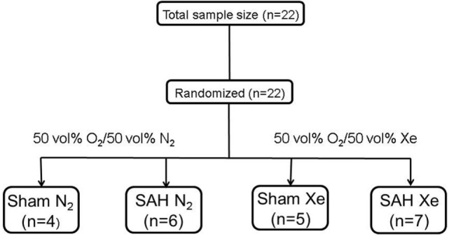
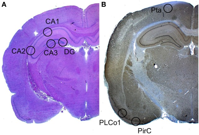
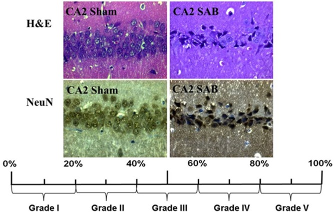
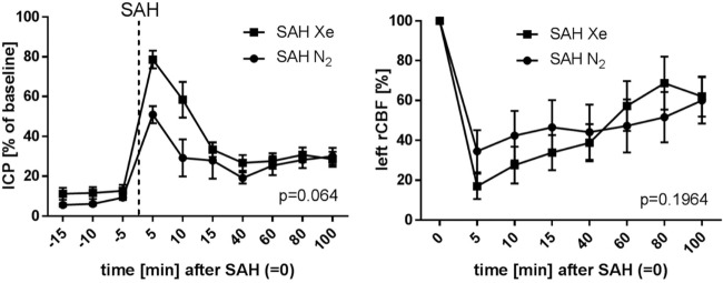
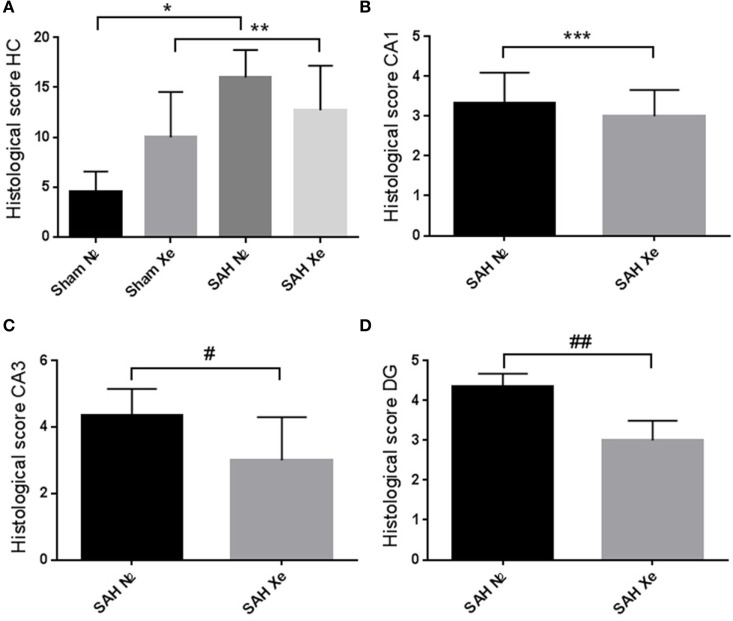
Sprague-Dawley rats ( = 22) were randomly assigned to receive either Sham surgery ( = 9; divided into two groups) or SAH induction endovascular perforation ( = 13, divided into two groups). Of those randomized for SAH, 7 animals were postoperatively ventilated with 50 vol% oxygen/50 vol% xenon for 1 h and 6 received 50 vol% oxygen/50 vol% nitrogen (control). The animals were sacrificed 24 h after SAH. Of each animal, a cerebral coronal section (-3.60 mm from bregma) was selected for assessment of histological damage 24 h after SAH. A 5-point neurohistopathological severity score was applied to assess neuronal cell damage in H&E and NeuN stained sections in a total of four predefined anatomical regions of interest. Microglial activation was evaluated by a software-assisted cell count of Iba-1 stained slices in three cortical regions of interest.
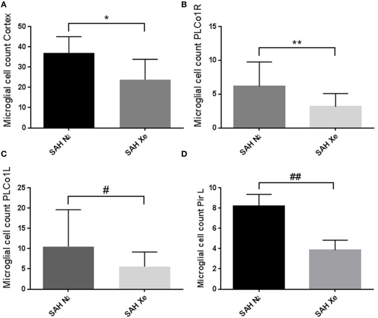
A diffuse cellular damage was apparent in all regions of the ipsilateral hippocampus 24 h after SAH. Xenon-treated animals presented with a milder damage after SAH. This effect was found to be particularly pronounced in the medial regions of the hippocampus, CA3 ( = 0.040), and dentate gyrus (DG = 0.040). However, for the CA1 and CA2 regions, there were no statistical differences in neuronal damage according to our histological scoring. A cell count of activated microglia was lower in the cortex of xenon-treated animals. This difference was especially apparent in the left piriform cortex ( = 0.017).
In animals treated with 50 vol% xenon (for 1 h) after SAH, a less pronounced neuronal damage was observed for the ipsilateral hippocampal regions CA3 and DG, when compared to the control group. In xenon-treated animals, a lower microglial cell count was observed suggesting an immunomodulatory effect generated by xenon. As for now, these results cannot be generalized as only some hippocampal regions are affected. Future studies should assess the time and localization dependency of xenon's beneficial properties after SAH.
稀有气体氙的神经保护特性已在多种损伤模型中得到证实。在此,我们首次在大鼠蛛网膜下腔出血(SAH)模型中研究氙在减轻早期脑损伤(EBI)方面的可能作用及其对出血后小胶质细胞神经炎症的影响。
将22只Sprague-Dawley大鼠随机分为接受假手术组(n = 9;分为两组)或SAH诱导组(通过血管内穿刺,n = 13,分为两组)。在随机接受SAH的大鼠中,7只动物术后用50体积%氧气/50体积%氙通气1小时,6只接受50体积%氧气/50体积%氮气(对照组)。SAH后24小时处死动物。从每只动物中选取距前囟-3.60毫米的脑冠状切片,用于评估SAH后24小时的组织学损伤。应用5分神经组织病理学严重程度评分,在苏木精-伊红(H&E)和NeuN染色切片中,对总共四个预定义的感兴趣解剖区域的神经元细胞损伤进行评估。通过软件辅助对三个感兴趣皮质区域的Iba-1染色切片进行细胞计数,评估小胶质细胞活化情况。
SAH后24小时,同侧海马体所有区域均出现弥漫性细胞损伤。氙处理组动物在SAH后损伤较轻。这种效应在海马体的内侧区域、CA3(p = 0.040)和齿状回(DG,p = 0.040)尤为明显。然而,对于CA1和CA2区域,根据我们的组织学评分,神经元损伤没有统计学差异。氙处理组动物皮质中活化小胶质细胞的细胞计数较低。这种差异在左侧梨状皮质尤为明显(p = 0.017)。
与对照组相比,SAH后用50体积%氙处理(1小时)的动物,同侧海马体区域CA3和DG的神经元损伤不太明显。在氙处理的动物中,观察到小胶质细胞计数较低,提示氙产生了免疫调节作用。目前,由于仅部分海马体区域受到影响,这些结果尚不能推广。未来的研究应评估SAH后氙有益特性的时间和定位依赖性。