Thomas Eloïse, Colombeau Ludovic, Gries Mickaël, Peterlini Thibaut, Mathieu Clélia, Thomas Noémie, Boura Cédric, Frochot Céline, Vanderesse Régis, Lux François, Barberi-Heyob Muriel, Tillement Olivier
Université Lyon, Université Claude Bernard Lyon 1, Centre National de la Recherche Scientifique (CNRS), Institut Lumière Matière, Lyon.
Laboratoire Réactions et Génie des Procédés, Université de Lorraine-CNRS, Nancy.
Int J Nanomedicine. 2017 Sep 26;12:7075-7088. doi: 10.2147/IJN.S141559. eCollection 2017.
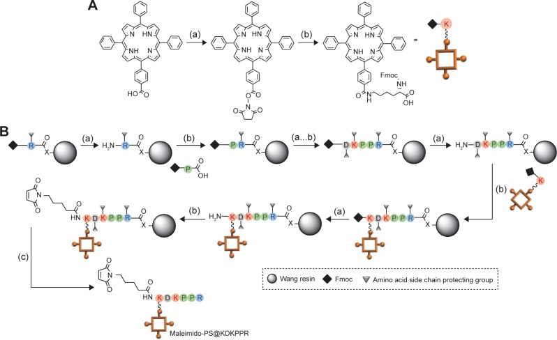
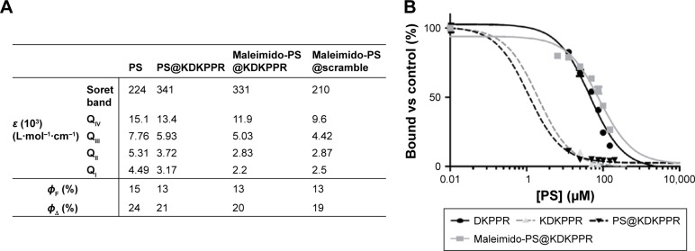
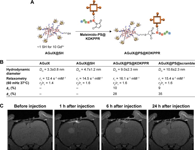
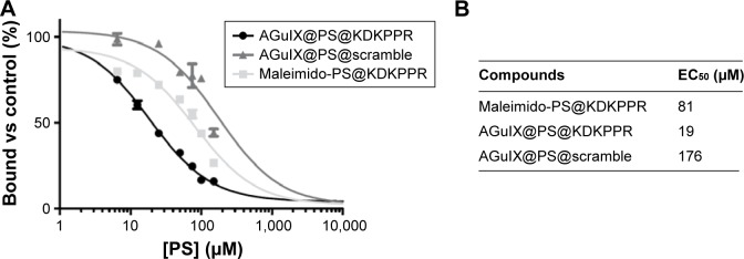
Despite combined treatments, glioblastoma outcome remains poor with frequent local recurrences, indicating that a more efficient and local therapy is needed. In this way, vascular-targeted photodynamic therapy (VTP) could help tumor eradication by destroying its neovessels. In this study, we designed a polysiloxane-based nanoparticle (NP) combining a magnetic resonance imaging (MRI) contrast agent, a photosensitizer (PS) and a new ligand peptide motif (KDKPPR) targeting neuropilin-1 (NRP-1), a receptor overexpressed by angiogenic endothelial cells of the tumor vasculature. This structure achieves the detection of the tumor tissue and its proliferating part by MRI analysis, followed by its treatment by VTP. The photophysical properties of the PS and the peptide affinity for NRP-1 recombinant protein were preserved after the functionalization of NPs. Cellular uptake of NPs by human umbilical vein endothelial cells (HUVEC) was increased twice compared to NPs without the KDKPPR peptide moiety or conjugated with a scramble peptide. NPs induced no cytotoxicity without light exposure but conferred a photocytotoxic effect to cells after photodynamic therapy (PDT). The in vivo selectivity, evaluated using a skinfold chamber model in mice, confirms that the functionalized NPs with KDKPPR peptide moiety were localized in the tumor vessel wall.
尽管采用了联合治疗,但胶质母细胞瘤的预后仍然很差,局部复发频繁,这表明需要一种更有效且局部的治疗方法。通过这种方式,血管靶向光动力疗法(VTP)可以通过破坏肿瘤的新生血管来帮助根除肿瘤。在本研究中,我们设计了一种基于聚硅氧烷的纳米颗粒(NP),它结合了磁共振成像(MRI)造影剂、光敏剂(PS)和一种靶向神经纤毛蛋白-1(NRP-1)的新配体肽基序(KDKPPR),NRP-1是肿瘤脉管系统中血管生成内皮细胞过度表达的一种受体。这种结构通过MRI分析实现对肿瘤组织及其增殖部分的检测,随后通过VTP进行治疗。在NP功能化后,PS的光物理性质以及肽对NRP-1重组蛋白的亲和力得以保留。与没有KDKPPR肽部分或与乱序肽偶联的NP相比,人脐静脉内皮细胞(HUVEC)对NP的细胞摄取增加了两倍。NP在无光照时不诱导细胞毒性,但在光动力疗法(PDT)后对细胞产生光细胞毒性作用。使用小鼠皮褶腔室模型评估的体内选择性证实,带有KDKPPR肽部分的功能化NP定位于肿瘤血管壁。