McGarry Matthew, Nauleau Pierre, Apostolakis Iason, Konofagou Elisa
Department of Biomedical Engineering, Columbia University, New York, NY, United States; Thayer School of Engineering, Dartmouth College, Hanover, NH, United States.
Department of Biomedical Engineering, Columbia University, New York, NY, United States.
J Biomech. 2017 Nov 7;64:136-144. doi: 10.1016/j.jbiomech.2017.09.017. Epub 2017 Sep 27.
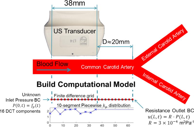
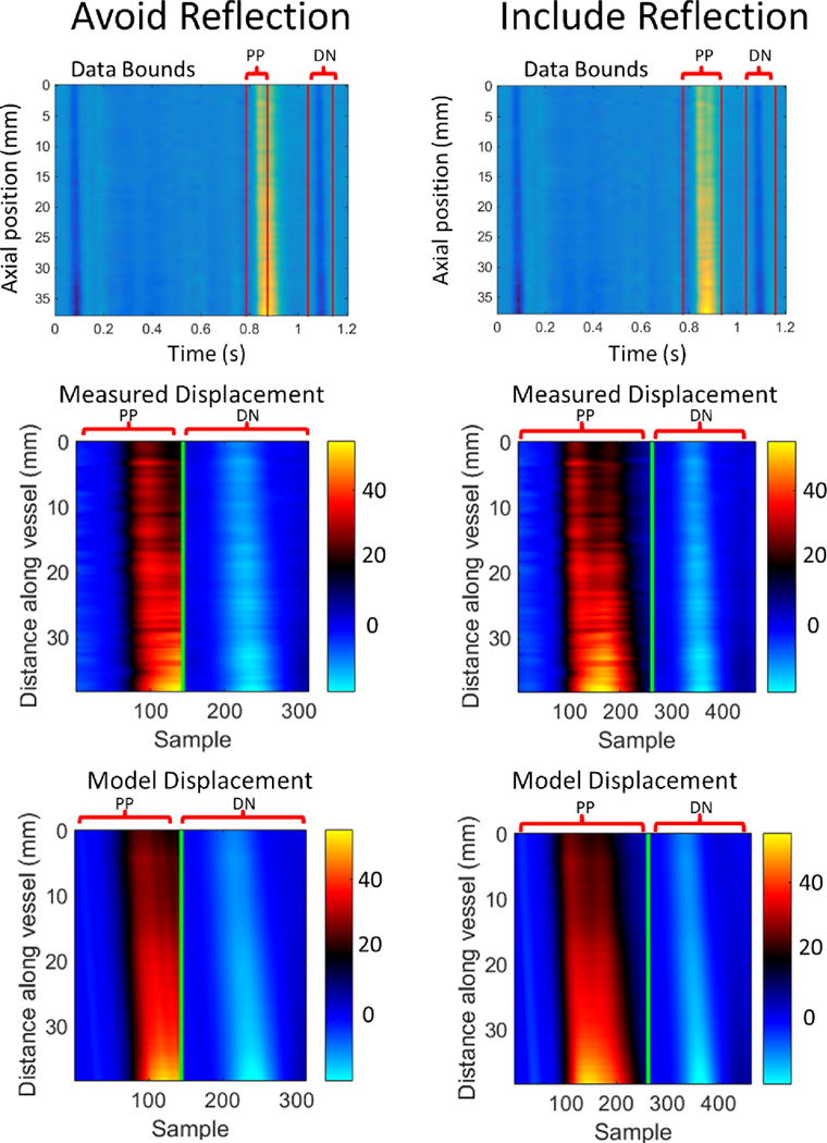
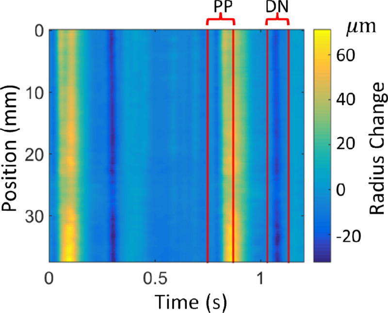
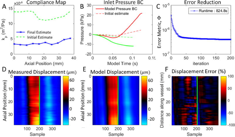
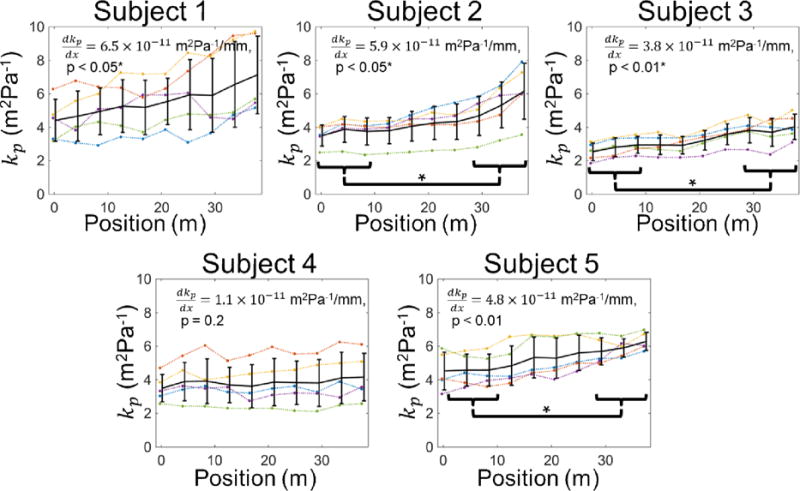
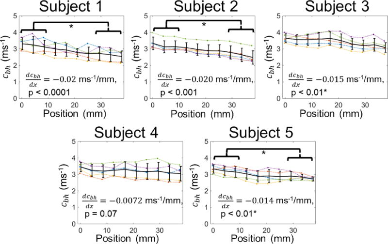
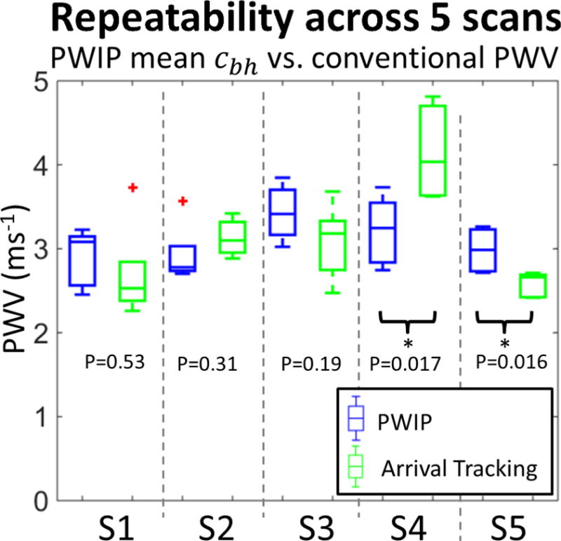
Accurate arterial stiffness measurement would improve diagnosis and monitoring for many diseases. Atherosclerotic plaques and aneurysms are expected to involve focal changes in vessel wall properties; therefore, a method to image the stiffness variation would be a valuable clinical tool. The pulse wave inverse problem (PWIP) fits unknown parameters from a computational model of arterial pulse wave propagation to ultrasound-based measurements of vessel wall displacements by minimizing the difference between the model and measured displacements. The PWIP has been validated in phantoms, and this study presents the first in vivo demonstration. The common carotid arteries of five healthy volunteers were imaged five times in a single session with repositioning of the probe and subject between each scan. The 1D finite difference computational model used in the PWIP spanned from the start of the transducer to the carotid bifurcation, where a resistance outlet boundary condition was applied to approximately model the downstream reflection of the pulse wave. Unknown parameters that were estimated by the PWIP included a 10-segment linear piecewise compliance distribution and 16 discrete cosine transformation coefficients for each of the inlet boundary conditions. Input data was selected to include pulse waves resulting from the primary pulse and dicrotic notch. The recovered compliance maps indicate that the compliance increases close to the bifurcation, and the variability of the average pulse wave velocity estimated through the PWIP is on the order of 11%, which is similar to that of the conventional processing technique which tracks the wavefront arrival time (13%).
准确测量动脉僵硬度将改善多种疾病的诊断和监测。动脉粥样硬化斑块和动脉瘤预计会涉及血管壁特性的局部变化;因此,一种能够成像僵硬度变化的方法将是一种有价值的临床工具。脉搏波逆问题(PWIP)通过最小化模型与测量位移之间的差异,将动脉脉搏波传播计算模型中的未知参数与基于超声的血管壁位移测量值进行拟合。PWIP已在模型中得到验证,本研究首次进行了体内演示。在一次检查中,对五名健康志愿者的颈总动脉进行了五次成像,每次扫描之间重新定位探头和受试者。PWIP中使用的一维有限差分计算模型从换能器起点延伸至颈动脉分叉处,在该分叉处应用了阻力出口边界条件,以近似模拟脉搏波的下游反射。由PWIP估计的未知参数包括一个10段线性分段顺应性分布以及每个入口边界条件的16个离散余弦变换系数。选择的输入数据包括由主脉冲和重搏波切迹产生的脉搏波。恢复的顺应性图表明,靠近分叉处顺应性增加,通过PWIP估计的平均脉搏波速度的变异性约为11%,这与跟踪波前到达时间的传统处理技术的变异性(13%)相似。