Institute of Cognitive Neuroscience, University College London, London, UK.
Division of Language and Communication Science, City University London, London, UK.
Brain. 2017 Nov 1;140(11):3039-3054. doi: 10.1093/brain/awx234.
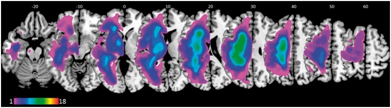
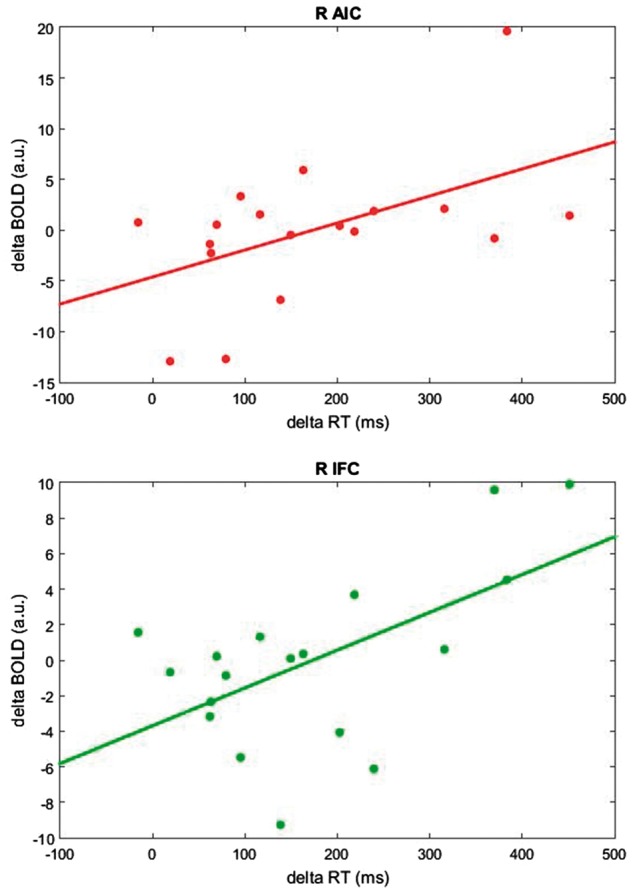
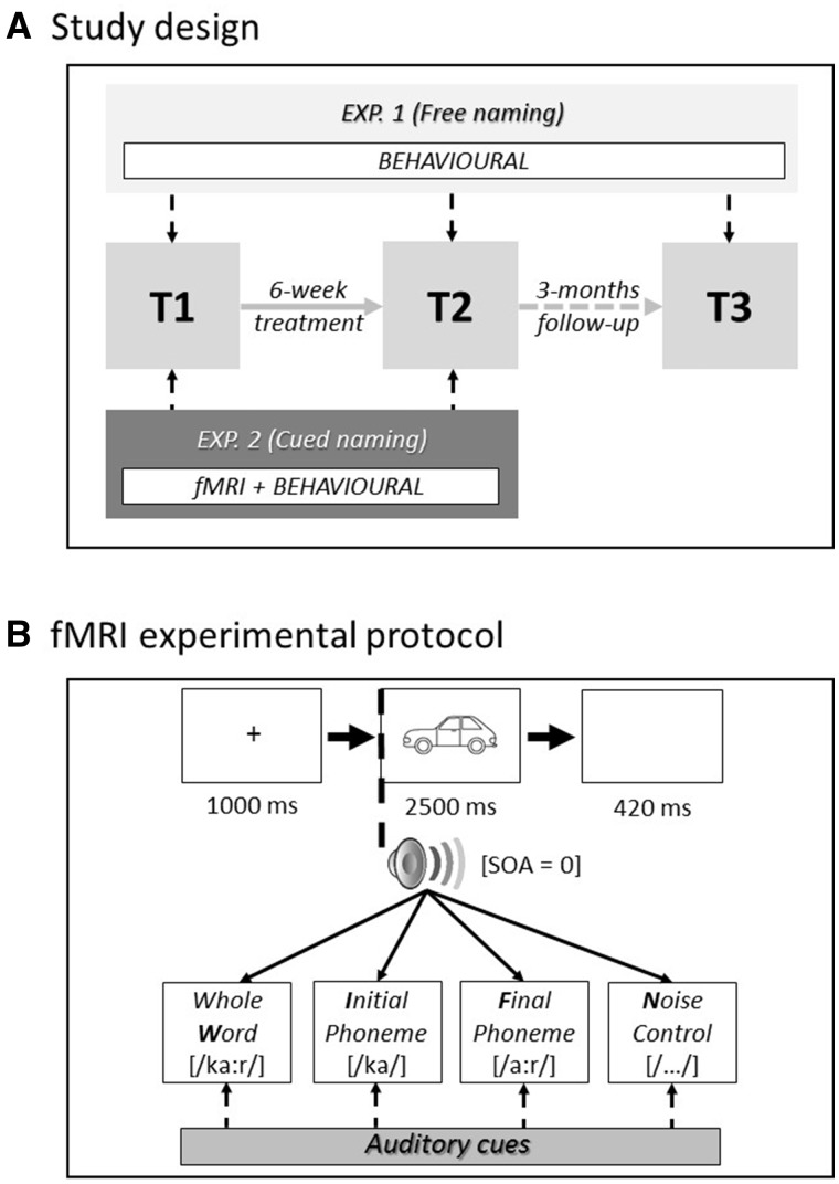
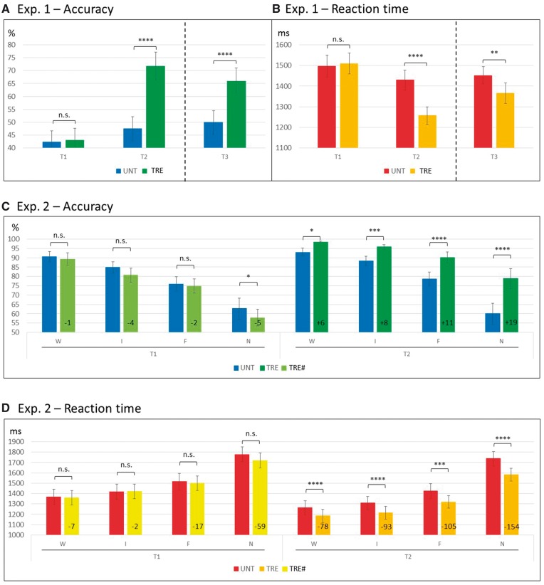
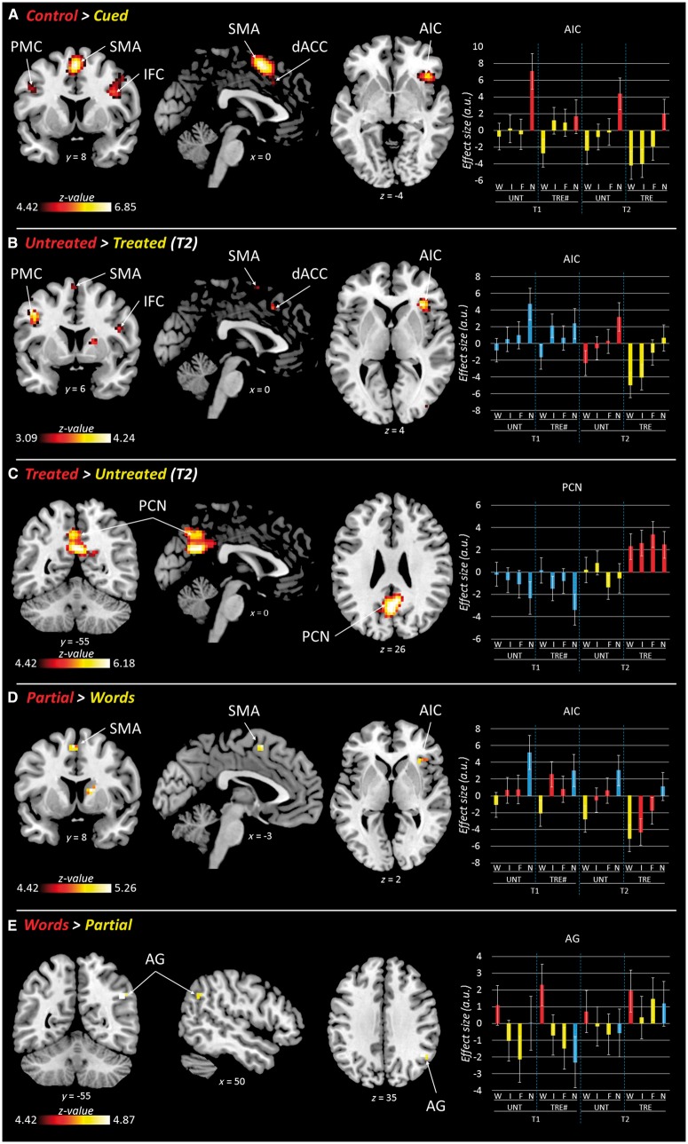
See Thompson and Woollams (doi:10.1093/brain/awx264) for a scientific commentary on this article. Previous research with aphasic patients has shown that picture naming can be facilitated by concurrent phonemic cueing [e.g. initial phoneme(s) of the word that the patient is trying to retrieve], both as an immediate word retrieval technique, and when practiced repeatedly over time as a long-term anomia treatment. Here, to investigate the neural mechanisms supporting word retrieval, we adopted—for the first time—a functional magnetic resonance imaging task using the same naming procedure as it occurs during the anomia treatment process. Before and directly after a 6-week anomia treatment programme, 18 chronic aphasic stroke patients completed our functional magnetic resonance imaging protocol—a picture naming task aided by three different types of phonemic cues (whole words, initial phonemes, final phonemes) and a noise-control condition. Patients completed a naming task based on the training materials, and a more general comprehensive battery of language tests both before and after the anomia treatment, to determine the effectiveness and specificity of the therapy. Our results demonstrate that the anomia treatment was effective and specific to speech production, significantly improving both patients’ naming accuracy and reaction time immediately post-treatment (unstandardized effect size: 29% and 17%, respectively; Cohen’s d: 3.45 and 1.83). Longer term gains in naming were maintained 3 months later. Functional imaging results showed that both immediate and long-term facilitation of naming involved a largely overlapping bilateral frontal network including the right anterior insula, inferior frontal and dorsal anterior cingulate cortices, and the left premotor cortex. These areas were associated with a neural priming effect (i.e. reduced blood oxygen level-dependent signal) during both immediate (phonemically-cued versus control-cue conditions), and long-term facilitation of naming (i.e. treated versus untreated items). Of note is that different brain regions were sensitive to different phonemic cue types. Processing of whole word cues was associated with increased activity in the right angular gyrus; whereas partial word cues (initial and final phonemes) recruited the left supplementary motor area, and right anterior insula, inferior frontal cortex, and basal ganglia. The recruitment of multiple and bilateral areas may help explain why phonemic cueing is such a successful behavioural facilitation tool for anomia treatment. Our results have important implications for optimizing current anomia treatment approaches, developing new treatments, and improving speech outcome for aphasic patients.
请参阅 Thompson 和 Woollams(doi:10.1093/brain/awx264)的科学评论,了解本文的相关内容。先前针对失语症患者的研究表明,图片命名可以通过同时进行语音提示来促进[例如,患者试图检索的单词的首字母(s)],既可以作为即时单词检索技术,也可以在一段时间内重复练习作为长期命名障碍治疗。在这里,为了研究支持单词检索的神经机制,我们首次采用功能磁共振成像任务,使用与命名障碍治疗过程中相同的命名程序。在进行为期 6 周的命名障碍治疗计划之前和之后,18 名慢性失语症中风患者完成了我们的功能磁共振成像协议 - 图片命名任务,辅助使用三种不同类型的语音提示(完整单词、首字母、尾字母)和噪音控制条件。患者根据训练材料完成命名任务,并在命名障碍治疗前后完成更全面的综合语言测试,以确定治疗的有效性和特异性。我们的结果表明,命名障碍治疗是有效的,并且对言语生成具有特异性,可显著提高患者治疗后即刻的命名准确性和反应时间(未标准化效应大小:分别为 29%和 17%;Cohen 的 d:3.45 和 1.83)。3 个月后,命名的长期收益得以维持。功能成像结果表明,命名的即时和长期促进都涉及一个基本重叠的双侧额网络,包括右侧前岛叶、下额和背侧前扣带皮层以及左侧运动前皮层。这些区域与神经启动效应(即依赖血氧水平的信号减少)相关,无论是在即时(语音提示与对照提示条件)还是长期促进命名(即治疗与未治疗项目)。值得注意的是,不同的大脑区域对不同的语音提示类型敏感。处理完整单词提示会引起右侧角回的活动增加;而部分单词提示(首字母和尾字母)会募集左侧辅助运动区、右侧前岛叶、下额和基底节。多个和双侧区域的募集可能有助于解释为什么语音提示是命名障碍治疗如此成功的行为促进工具。我们的结果对优化当前的命名障碍治疗方法、开发新的治疗方法以及改善失语症患者的言语预后具有重要意义。