• 文献检索
  • 文档翻译
  • 深度研究
  • 学术资讯
  • Suppr Zotero 插件Zotero 插件
  • 邀请有礼
  • 套餐&价格
  • 历史记录
应用&插件
Suppr Zotero 插件Zotero 插件浏览器插件Mac 客户端Windows 客户端微信小程序
定价
高级版会员购买积分包购买API积分包
服务
文献检索文档翻译深度研究API 文档MCP 服务
关于我们
关于 Suppr公司介绍联系我们用户协议隐私条款
关注我们

Suppr 超能文献

核心技术专利:CN118964589B侵权必究
粤ICP备2023148730 号-1Suppr @ 2026

文献检索

告别复杂PubMed语法,用中文像聊天一样搜索,搜遍4000万医学文献。AI智能推荐,让科研检索更轻松。

立即免费搜索

文件翻译

保留排版,准确专业,支持PDF/Word/PPT等文件格式,支持 12+语言互译。

免费翻译文档

深度研究

AI帮你快速写综述,25分钟生成高质量综述,智能提取关键信息,辅助科研写作。

立即免费体验

依赖于中心体的微管和肌动蛋白动力学调控中心体的保留和神经元的分层。

Inter-dependent apical microtubule and actin dynamics orchestrate centrosome retention and neuronal delamination.

机构信息

Division of Cell and Developmental Biology, School of Life Sciences, University of Dundee, Dundee, United Kingdom.

出版信息

Elife. 2017 Oct 23;6:e26215. doi: 10.7554/eLife.26215.

DOI:10.7554/eLife.26215
PMID:29058679
原文链接:https://pmc.ncbi.nlm.nih.gov/articles/PMC5653239/
Abstract

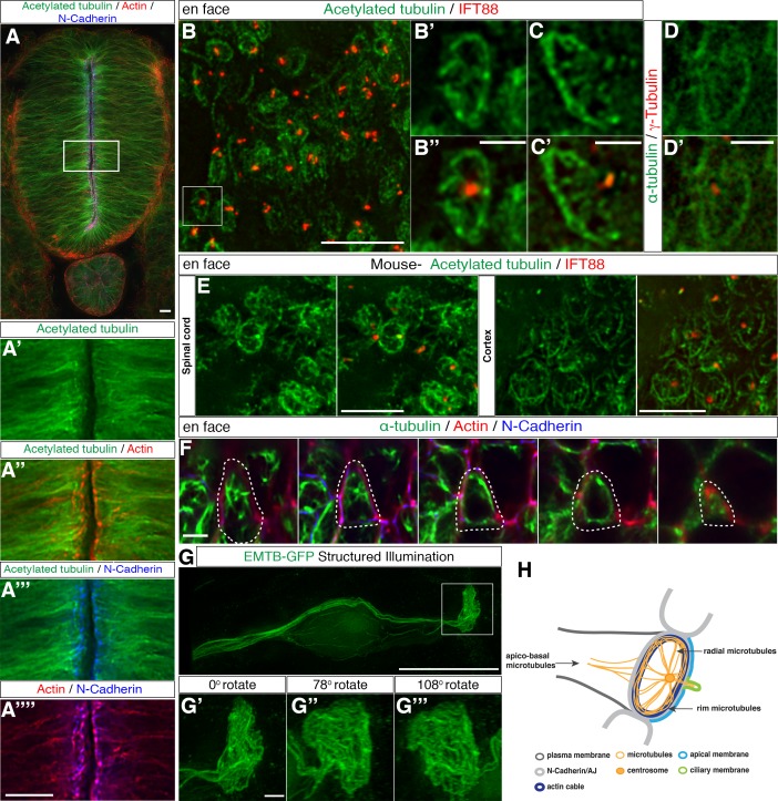
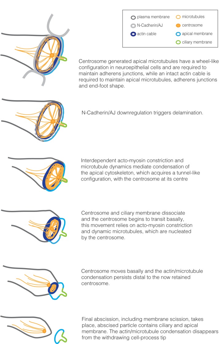
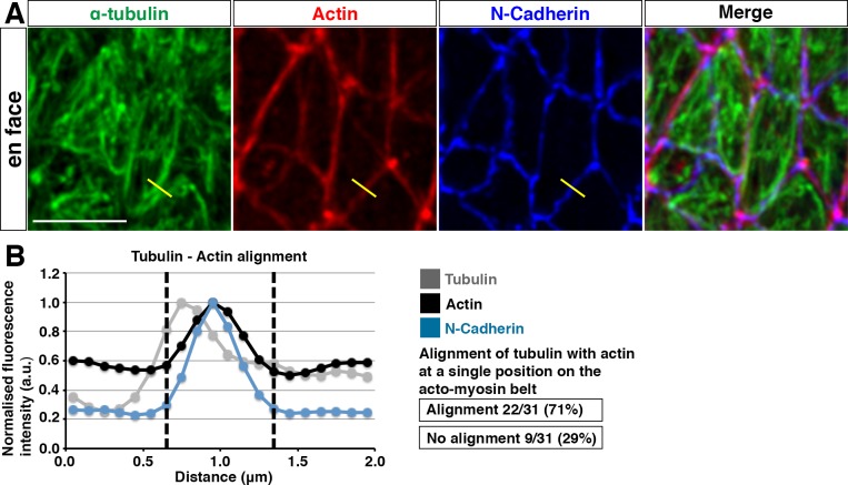
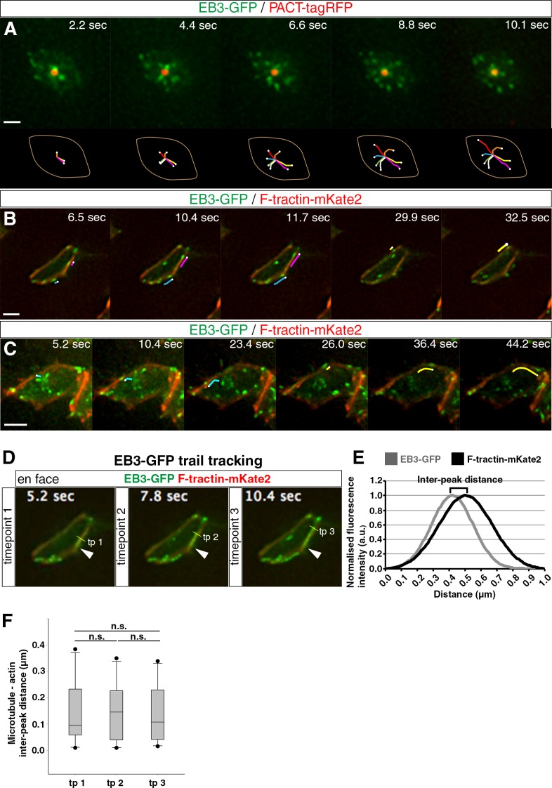
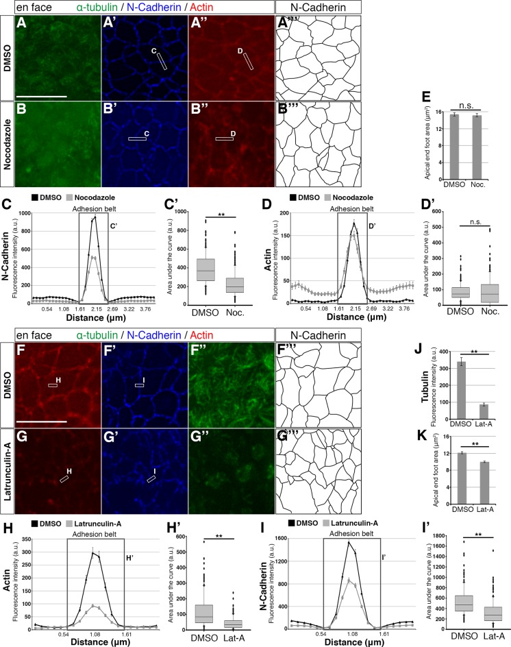
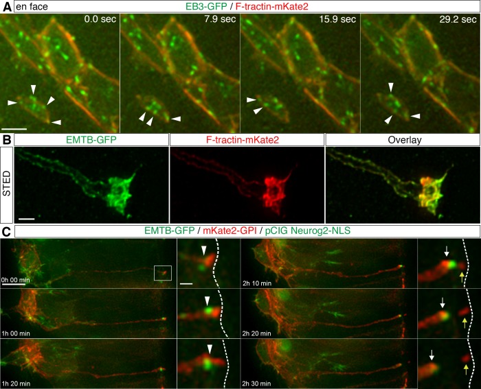
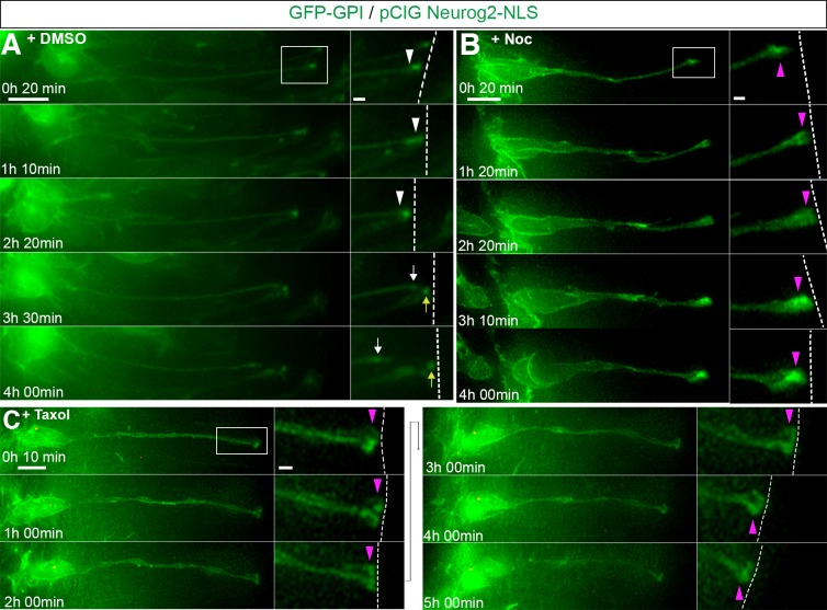
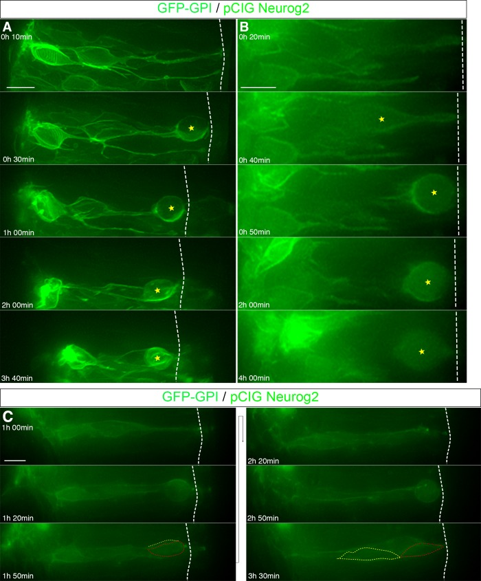
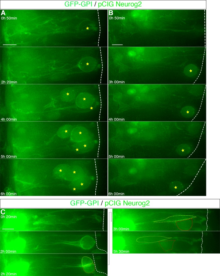
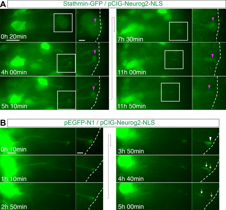
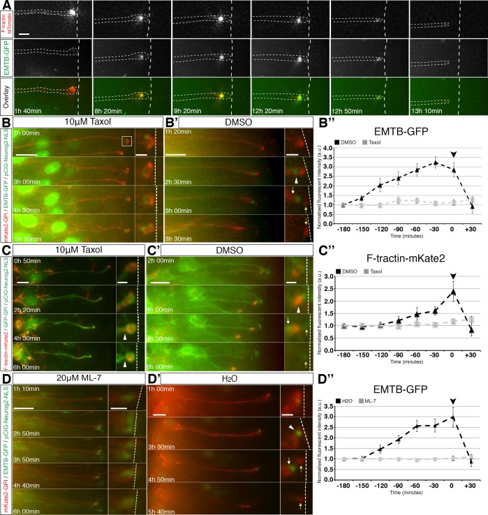
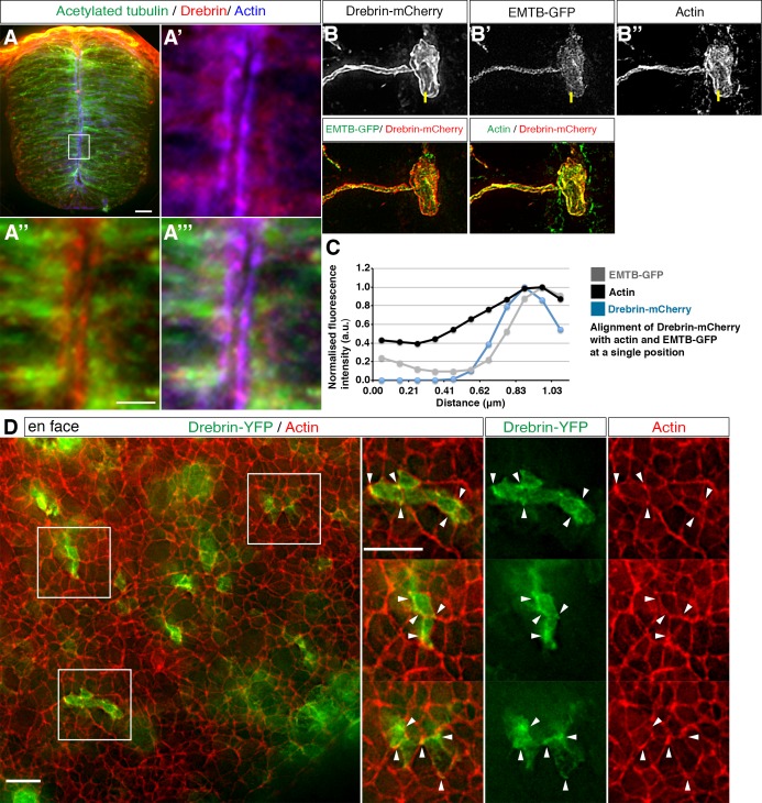
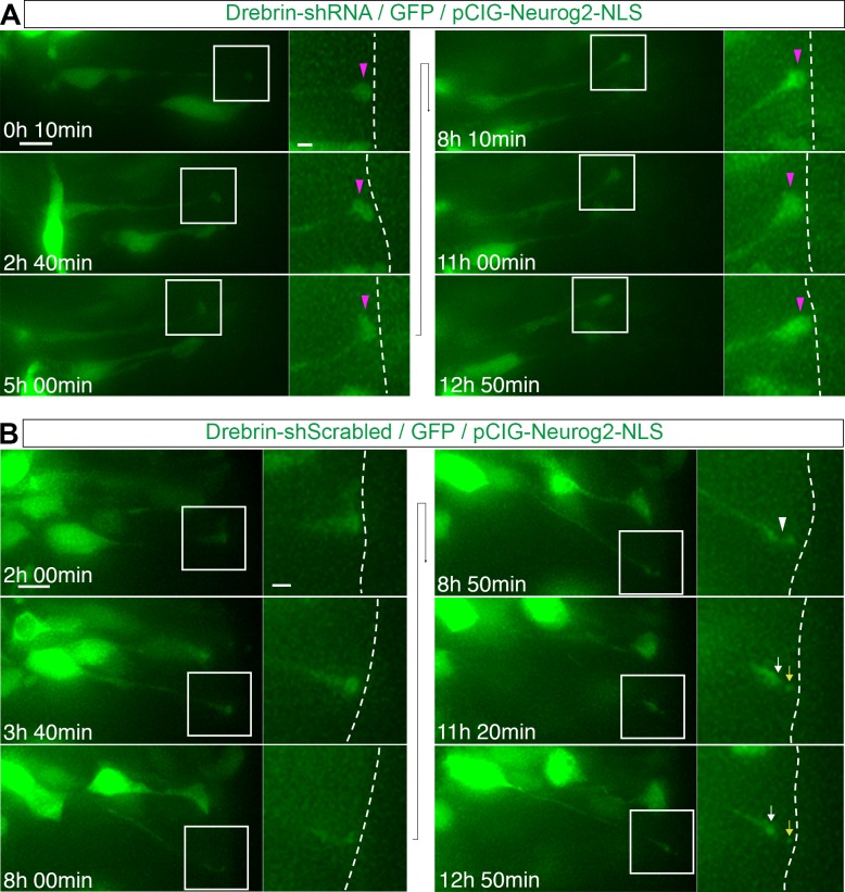
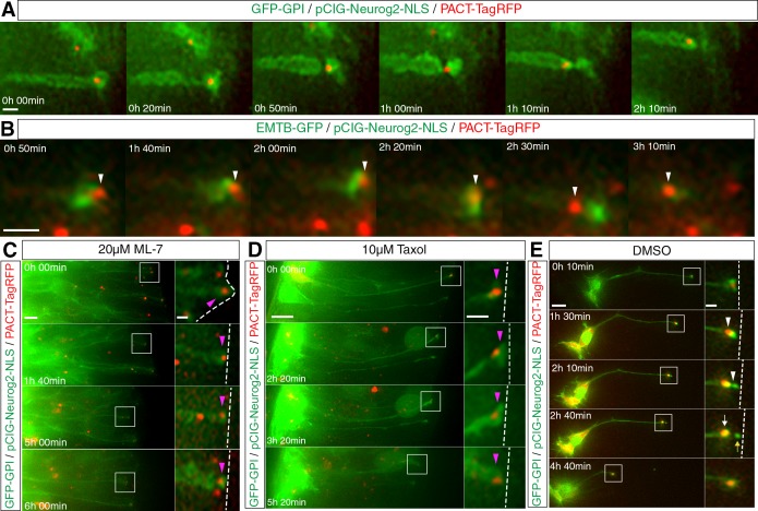
Detachment of newborn neurons from the neuroepithelium is required for correct neuronal architecture and functional circuitry. This process, also known as delamination, involves adherens-junction disassembly and acto-myosin-mediated abscission, during which the centrosome is retained while apical/ciliary membranes are shed. Cell-biological mechanisms mediating delamination are, however, poorly understood. Using live-tissue and super-resolution imaging, we uncover a centrosome-nucleated wheel-like microtubule configuration, aligned with the apical actin cable and adherens-junctions within chick and mouse neuroepithelial cells. These microtubules maintain adherens-junctions while actin maintains microtubules, adherens-junctions and apical end-foot dimensions. During neuronal delamination, acto-myosin constriction generates a tunnel-like actin-microtubule configuration through which the centrosome translocates. This movement requires inter-dependent actin and microtubule activity, and we identify drebrin as a potential coordinator of these cytoskeletal dynamics. Furthermore, centrosome compromise revealed that this organelle is required for delamination. These findings identify new cytoskeletal configurations and regulatory relationships that orchestrate neuronal delamination and may inform mechanisms underlying pathological epithelial cell detachment.

摘要

新生儿神经元从神经上皮分离对于正确的神经元结构和功能回路是必需的。这个过程也称为分层,涉及黏着连接的解体和肌动球蛋白介导的断裂,在此过程中,中心体被保留,而顶/纤毛膜被丢弃。然而,介导分层的细胞生物学机制还知之甚少。使用活体组织和超分辨率成像,我们在鸡和鼠神经上皮细胞中发现了一种由中心体引发的轮状微管结构,与顶端肌动蛋白电缆和黏着连接对齐。这些微管在维持黏着连接的同时,肌动蛋白维持微管、黏着连接和顶端足的尺寸。在神经元分层过程中,肌球蛋白收缩产生一种隧道样的肌动蛋白微管结构,中心体通过该结构迁移。这种运动需要依赖肌动蛋白和微管的活性,我们确定 drebrin 是这些细胞骨架动力学的潜在协调蛋白。此外,中心体受损表明该细胞器对于分层是必需的。这些发现确定了新的细胞骨架结构和调节关系,这些关系协调神经元分层,并可能为上皮细胞病理性分离的机制提供信息。

https://cdn.ncbi.nlm.nih.gov/pmc/blobs/e413/5653239/21c1aa07d1be/elife-26215-fig10.jpg
https://cdn.ncbi.nlm.nih.gov/pmc/blobs/e413/5653239/1972106439c5/elife-26215-fig1.jpg
https://cdn.ncbi.nlm.nih.gov/pmc/blobs/e413/5653239/bc077c63982d/elife-26215-fig1-figsupp1.jpg
https://cdn.ncbi.nlm.nih.gov/pmc/blobs/e413/5653239/1efb65b5aab4/elife-26215-fig2.jpg
https://cdn.ncbi.nlm.nih.gov/pmc/blobs/e413/5653239/c1defc83cb02/elife-26215-fig3.jpg
https://cdn.ncbi.nlm.nih.gov/pmc/blobs/e413/5653239/d1b37202bd64/elife-26215-fig4.jpg
https://cdn.ncbi.nlm.nih.gov/pmc/blobs/e413/5653239/fa97ec463828/elife-26215-fig5.jpg
https://cdn.ncbi.nlm.nih.gov/pmc/blobs/e413/5653239/9d0686f3ff29/elife-26215-fig5-figsupp1.jpg
https://cdn.ncbi.nlm.nih.gov/pmc/blobs/e413/5653239/d8f5849aa063/elife-26215-fig5-figsupp2.jpg
https://cdn.ncbi.nlm.nih.gov/pmc/blobs/e413/5653239/0a6289c0654c/elife-26215-fig5-figsupp3.jpg
https://cdn.ncbi.nlm.nih.gov/pmc/blobs/e413/5653239/494229da8168/elife-26215-fig6.jpg
https://cdn.ncbi.nlm.nih.gov/pmc/blobs/e413/5653239/56f8f9956063/elife-26215-fig7.jpg
https://cdn.ncbi.nlm.nih.gov/pmc/blobs/e413/5653239/208a761f36b3/elife-26215-fig7-figsupp1.jpg
https://cdn.ncbi.nlm.nih.gov/pmc/blobs/e413/5653239/a09d0d090343/elife-26215-fig8.jpg
https://cdn.ncbi.nlm.nih.gov/pmc/blobs/e413/5653239/21c1aa07d1be/elife-26215-fig10.jpg

相似文献

1
Inter-dependent apical microtubule and actin dynamics orchestrate centrosome retention and neuronal delamination.依赖于中心体的微管和肌动蛋白动力学调控中心体的保留和神经元的分层。
Elife. 2017 Oct 23;6:e26215. doi: 10.7554/eLife.26215.
2
Microtubules promote intercellular contractile force transmission during tissue folding.微管促进组织折叠过程中的细胞间收缩力传递。
J Cell Biol. 2019 Aug 5;218(8):2726-2742. doi: 10.1083/jcb.201902011. Epub 2019 Jun 21.
3
Cell biological mechanisms regulating chick neurogenesis.调节鸡神经发生的细胞生物学机制。
Int J Dev Biol. 2018;62(1-2-3):167-175. doi: 10.1387/ijdb.170268ks.
4
Neuronal Delamination and Outer Radial Glia Generation in Neocortical Development.新皮层发育中的神经元脱层与外放射状胶质细胞生成
Front Cell Dev Biol. 2021 Feb 5;8:623573. doi: 10.3389/fcell.2020.623573. eCollection 2020.
5
Actin filaments regulate microtubule growth at the centrosome.肌动蛋白丝调节中心体处的微管生长。
EMBO J. 2019 Jun 3;38(11). doi: 10.15252/embj.201899630. Epub 2019 Mar 22.
6
A release-and-capture mechanism generates an essential non-centrosomal microtubule array during tube budding.释出-捕获机制在管状芽生过程中产生一个必需的非中心体微管阵列。
Nat Commun. 2021 Jul 2;12(1):4096. doi: 10.1038/s41467-021-24332-0.
7
Acto-myosin network geometry defines centrosome position.肌球蛋白网络的几何形状决定了中心体的位置。
Curr Biol. 2021 Mar 22;31(6):1206-1220.e5. doi: 10.1016/j.cub.2021.01.002. Epub 2021 Feb 19.
8
Dictyostelium LIS1 is a centrosomal protein required for microtubule/cell cortex interactions, nucleus/centrosome linkage, and actin dynamics.盘基网柄菌LIS1是一种中心体蛋白,对于微管/细胞皮层相互作用、细胞核/中心体连接以及肌动蛋白动力学而言是必需的。
Mol Biol Cell. 2005 Jun;16(6):2759-71. doi: 10.1091/mbc.e05-01-0069. Epub 2005 Mar 30.
9
Ninein is released from the centrosome and moves bi-directionally along microtubules.九蛋白从中心体释放,并沿微管双向移动。
J Cell Sci. 2007 Sep 1;120(Pt 17):3064-74. doi: 10.1242/jcs.010322. Epub 2007 Aug 14.
10
Radial somatic F-actin organization affects growth cone dynamics during early neuronal development.径向体 F-肌动蛋白组织影响早期神经元发育过程中的生长锥动力学。
EMBO Rep. 2019 Dec 5;20(12):e47743. doi: 10.15252/embr.201947743. Epub 2019 Oct 24.

引用本文的文献

1
Early spinal cord development: from neural tube formation to neurogenesis.早期脊髓发育:从神经管形成到神经发生
Nat Rev Neurosci. 2025 Apr;26(4):195-213. doi: 10.1038/s41583-025-00906-5. Epub 2025 Feb 6.
2
Establishing neuronal polarity: microtubule regulation during neurite initiation.建立神经元极性:轴突起始过程中的微管调节
Oxf Open Neurosci. 2022 May 13;1:kvac007. doi: 10.1093/oons/kvac007. eCollection 2022.
3
Pioneer statoacoustic neurons guide neuroblast behaviour during otic ganglion assembly.先驱性的前庭听觉神经元在耳神经节装配过程中引导神经母细胞的行为。

本文引用的文献

1
Drebrin-mediated microtubule-actomyosin coupling steers cerebellar granule neuron nucleokinesis and migration pathway selection.Drebrin 通过介导微管-肌动蛋白偶联来控制小脑颗粒神经元的核迁移,并选择迁移路径。
Nat Commun. 2017 Feb 23;8:14484. doi: 10.1038/ncomms14484.
2
Polarity Reversal by Centrosome Repositioning Primes Cell Scattering during Epithelial-to-Mesenchymal Transition.通过中心体重新定位实现的极性反转引发上皮-间质转化过程中的细胞散射。
Dev Cell. 2017 Jan 23;40(2):168-184. doi: 10.1016/j.devcel.2016.12.004. Epub 2016 Dec 29.
3
Microtubule organization is determined by the shape of epithelial cells.
Development. 2023 Nov 1;150(21). doi: 10.1242/dev.201824. Epub 2023 Nov 8.
4
Vinculin is required for interkinetic nuclear migration (INM) and cell cycle progression.着丝粒蛋白对于核周运动(INM)和细胞周期进程是必需的。
J Cell Biol. 2024 Jan 1;223(1). doi: 10.1083/jcb.202106169. Epub 2023 Oct 27.
5
Asymmetric inheritance of centrosomes maintains stem cell properties in human neural progenitor cells.中心体的非对称遗传维持了人类神经祖细胞的干细胞特性。
Elife. 2023 Oct 26;12:e83157. doi: 10.7554/eLife.83157.
6
Connecting neurodevelopment to neurodegeneration: a spotlight on the role of kinesin superfamily protein 2A (KIF2A).连接神经发育与神经退行性变:聚焦驱动蛋白超家族蛋白2A(KIF2A)的作用
Neural Regen Res. 2024 Feb;19(2):375-379. doi: 10.4103/1673-5374.375298.
7
An actin filament branching surveillance system regulates cell cycle progression, cytokinesis and primary ciliogenesis.肌动蛋白丝分支监测系统调控细胞周期进程、胞质分裂和初级纤毛发生。
Nat Commun. 2023 Mar 27;14(1):1687. doi: 10.1038/s41467-023-37340-z.
8
Moonlighting at the Poles: Non-Canonical Functions of Centrosomes.两极的兼职:中心体的非经典功能
Front Cell Dev Biol. 2022 Jul 14;10:930355. doi: 10.3389/fcell.2022.930355. eCollection 2022.
9
The Symmetry of Neural Stem Cell and Progenitor Divisions in the Vertebrate Brain.脊椎动物大脑中神经干细胞和祖细胞分裂的对称性
Front Cell Dev Biol. 2022 May 25;10:885269. doi: 10.3389/fcell.2022.885269. eCollection 2022.
10
Regulation of Cell Delamination During Cortical Neurodevelopment and Implication for Brain Disorders.皮层神经发育过程中细胞脱层的调控及其对脑部疾病的影响
Front Neurosci. 2022 Feb 23;16:824802. doi: 10.3389/fnins.2022.824802. eCollection 2022.
微管组织由上皮细胞的形状决定。
Nat Commun. 2016 Oct 25;7:13172. doi: 10.1038/ncomms13172.
4
Neocortex expansion in development and evolution - from cell biology to single genes.大脑新皮层在发育和进化中的扩展——从细胞生物学到单个基因。
Curr Opin Neurobiol. 2016 Aug;39:122-32. doi: 10.1016/j.conb.2016.05.004. Epub 2016 May 31.
5
Zeb1 controls neuron differentiation and germinal zone exit by a mesenchymal-epithelial-like transition.Zeb1通过间充质-上皮样转变控制神经元分化和生发区迁出。
Elife. 2016 May 14;5:e12717. doi: 10.7554/eLife.12717.
6
Cadherin Trafficking for Tissue Morphogenesis: Control and Consequences.用于组织形态发生的钙黏蛋白运输:调控与影响
Traffic. 2016 Dec;17(12):1233-1243. doi: 10.1111/tra.12407. Epub 2016 May 19.
7
Characterization of primary cilia during the differentiation of retinal ganglion cells in the zebrafish.斑马鱼视网膜神经节细胞分化过程中初级纤毛的特征分析。
Neural Dev. 2016 Apr 6;11:10. doi: 10.1186/s13064-016-0064-z.
8
Coordinating neuronal actin-microtubule dynamics.协调神经元肌动蛋白-微管动力学。
Curr Biol. 2015 Aug 3;25(15):R677-91. doi: 10.1016/j.cub.2015.06.020.
9
Fat1 interacts with Fat4 to regulate neural tube closure, neural progenitor proliferation and apical constriction during mouse brain development.在小鼠大脑发育过程中,Fat1与Fat4相互作用,以调节神经管闭合、神经祖细胞增殖和顶端收缩。
Development. 2015 Aug 15;142(16):2781-91. doi: 10.1242/dev.123539. Epub 2015 Jul 24.
10
The role of adherens junctions in the developing neocortex.黏着连接在新皮质发育中的作用。
Cell Adh Migr. 2015;9(3):167-74. doi: 10.1080/19336918.2015.1027478.