• 文献检索
  • 文档翻译
  • 深度研究
  • 学术资讯
  • Suppr Zotero 插件Zotero 插件
  • 邀请有礼
  • 套餐&价格
  • 历史记录
应用&插件
Suppr Zotero 插件Zotero 插件浏览器插件Mac 客户端Windows 客户端微信小程序
定价
高级版会员购买积分包购买API积分包
服务
文献检索文档翻译深度研究API 文档MCP 服务
关于我们
关于 Suppr公司介绍联系我们用户协议隐私条款
关注我们

Suppr 超能文献

核心技术专利:CN118964589B侵权必究
粤ICP备2023148730 号-1Suppr @ 2026

文献检索

告别复杂PubMed语法,用中文像聊天一样搜索,搜遍4000万医学文献。AI智能推荐,让科研检索更轻松。

立即免费搜索

文件翻译

保留排版,准确专业,支持PDF/Word/PPT等文件格式,支持 12+语言互译。

免费翻译文档

深度研究

AI帮你快速写综述,25分钟生成高质量综述,智能提取关键信息,辅助科研写作。

立即免费体验

使用三次谐波产生显微镜对小鼠颅骨中的骨细胞进行活体成像。

Intravital imaging of osteocytes in mouse calvaria using third harmonic generation microscopy.

作者信息

Tokarz Danielle, Cisek Richard, Wein Marc N, Turcotte Raphaël, Haase Christa, Yeh Shu-Chi A, Bharadwaj Srinidhi, Raphael Anthony P, Paudel Hari, Alt Clemens, Liu Tzu-Ming, Kronenberg Henry M, Lin Charles P

机构信息

Advanced Microscopy Program, Center for Systems Biology and Wellman Center for Photomedicine, Massachusetts General Hospital, Harvard Medical School, Boston, Massachusetts, United States of America.

Department of Physical and Chemical Sciences, University of Toronto, Mississauga, Ontario, Canada.

出版信息

PLoS One. 2017 Oct 24;12(10):e0186846. doi: 10.1371/journal.pone.0186846. eCollection 2017.

DOI:10.1371/journal.pone.0186846
PMID:29065178
原文链接:https://pmc.ncbi.nlm.nih.gov/articles/PMC5655444/
Abstract

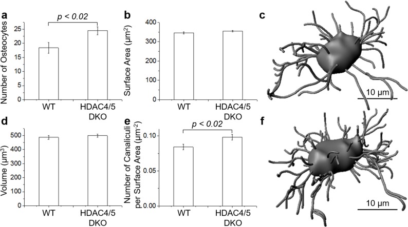
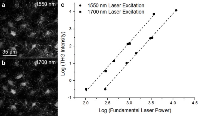
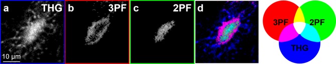
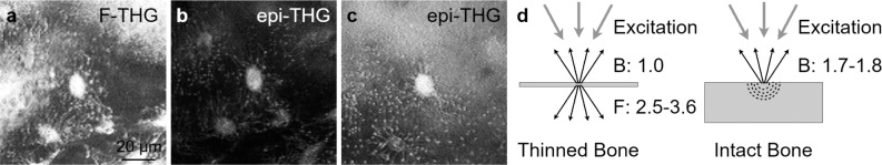
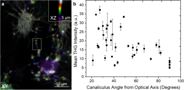
Osteocytes are the most abundant cell in the bone, and have multiple functions including mechanosensing and regulation of bone remodeling activities. Since osteocytes are embedded in the bone matrix, their inaccessibility makes in vivo studies problematic. Therefore, a non-invasive technique with high spatial resolution is desired. The purpose of this study is to investigate the use of third harmonic generation (THG) microscopy as a noninvasive technique for high-resolution imaging of the lacunar-canalicular network (LCN) in live mice. By performing THG imaging in combination with two- and three-photon fluorescence microscopy, we show that THG signal is produced from the bone-interstitial fluid boundary of the lacuna, while the interstitial fluid-osteocyte cell boundary shows a weaker THG signal. Canaliculi are also readily visualized by THG imaging, with canaliculi oriented at small angles relative to the optical axis exhibiting stronger signal intensity compared to those oriented perpendicular to the optical axis (parallel to the image plane). By measuring forward- versus epi-detected THG signals in thinned versus thick bone samples ex vivo, we found that the epi-collected THG from the LCN of intact bone contains a superposition of backward-directed and backscattered forward-THG. As an example of a biological application, THG was used as a label-free imaging technique to study structural variations in the LCN of live mice deficient in both histone deacetylase 4 and 5 (HDAC4, HDAC5). Three-dimensional analyses were performed and revealed statistically significant differences between the HDAC4/5 double knockout and wild type mice in the number of osteocytes per volume and the number of canaliculi per lacunar surface area. These changes in osteocyte density and dendritic projections occurred without differences in lacunar size. This study demonstrates that THG microscopy imaging of the LCN in live mice enables quantitative analysis of osteocytes in animal models without the use of dyes or physical sectioning.

摘要

骨细胞是骨骼中最丰富的细胞,具有多种功能,包括机械传感和调节骨重塑活动。由于骨细胞嵌入骨基质中,难以获取使得体内研究存在问题。因此,需要一种具有高空间分辨率的非侵入性技术。本研究的目的是探讨使用三次谐波产生(THG)显微镜作为一种非侵入性技术,用于对活小鼠的骨陷窝-骨小管网络(LCN)进行高分辨率成像。通过将THG成像与双光子和三光子荧光显微镜相结合,我们发现THG信号由骨陷窝的骨间质液边界产生,而间质液-骨细胞边界的THG信号较弱。骨小管也可通过THG成像清晰地可视化,相对于光轴呈小角度取向的骨小管比垂直于光轴(平行于图像平面)取向的骨小管表现出更强的信号强度。通过在体外对薄骨和厚骨样本中前向与背向检测的THG信号进行测量,我们发现从完整骨的LCN背向收集的THG包含向后定向和向前背向散射的THG叠加。作为一个生物学应用的例子,THG被用作一种无标记成像技术,以研究组蛋白脱乙酰酶4和5(HDAC4、HDAC5)均缺乏的活小鼠LCN中的结构变化。进行了三维分析,结果显示HDAC4/5双敲除小鼠和野生型小鼠在每体积骨细胞数量和每个骨陷窝表面积的骨小管数量上存在统计学显著差异。骨细胞密度和树突状突起的这些变化在骨陷窝大小上没有差异。本研究表明,对活小鼠的LCN进行THG显微镜成像能够在不使用染料或物理切片的情况下对动物模型中的骨细胞进行定量分析。

https://cdn.ncbi.nlm.nih.gov/pmc/blobs/1db7/5655444/32f0eec0b2a6/pone.0186846.g006.jpg
https://cdn.ncbi.nlm.nih.gov/pmc/blobs/1db7/5655444/3560ed28241e/pone.0186846.g001.jpg
https://cdn.ncbi.nlm.nih.gov/pmc/blobs/1db7/5655444/0084e62d64cc/pone.0186846.g002.jpg
https://cdn.ncbi.nlm.nih.gov/pmc/blobs/1db7/5655444/0b72e87ef4a2/pone.0186846.g003.jpg
https://cdn.ncbi.nlm.nih.gov/pmc/blobs/1db7/5655444/0b82f0923b6d/pone.0186846.g004.jpg
https://cdn.ncbi.nlm.nih.gov/pmc/blobs/1db7/5655444/679f24c09eaf/pone.0186846.g005.jpg
https://cdn.ncbi.nlm.nih.gov/pmc/blobs/1db7/5655444/32f0eec0b2a6/pone.0186846.g006.jpg

相似文献

1
Intravital imaging of osteocytes in mouse calvaria using third harmonic generation microscopy.使用三次谐波产生显微镜对小鼠颅骨中的骨细胞进行活体成像。
PLoS One. 2017 Oct 24;12(10):e0186846. doi: 10.1371/journal.pone.0186846. eCollection 2017.
2
Visualizing the "sandwich" structure of osteocytes in their native environment deep in bone in vivo.在骨内的天然环境中对活体内骨细胞的“三明治”结构进行可视化。
J Biophotonics. 2019 Apr;12(4):e201800360. doi: 10.1002/jbio.201800360. Epub 2018 Dec 13.
3
Alterations in the osteocyte lacunar-canalicular microenvironment due to estrogen deficiency.由于雌激素缺乏导致的破骨细胞陷窝-小管微环境的改变。
Bone. 2012 Sep;51(3):488-97. doi: 10.1016/j.bone.2012.05.014. Epub 2012 May 23.
4
NFATc1 Is Required for Vitamin D- and Phosphate-Mediated Regulation of Osteocyte Lacuno-Canalicular Remodeling.维生素D和磷酸盐介导的骨细胞陷窝-小管重塑调节需要NFATc1
Endocrinology. 2024 Jul 1;165(8). doi: 10.1210/endocr/bqae087.
5
The contribution of the pericanalicular matrix to mineral content in human osteonal bone.管周基质对人骨板层骨矿含量的贡献。
Bone. 2019 Jun;123:76-85. doi: 10.1016/j.bone.2019.03.018. Epub 2019 Mar 18.
6
Impaired 1,25 dihydroxyvitamin D3 action and hypophosphatemia underlie the altered lacuno-canalicular remodeling observed in the Hyp mouse model of XLH.XLH 模型鼠的骨小梁腔隙改建异常,其原因是 1,25 二羟维生素 D3 作用受损和低血磷。
PLoS One. 2021 May 27;16(5):e0252348. doi: 10.1371/journal.pone.0252348. eCollection 2021.
7
Disrupted osteocyte connectivity and pericellular fluid flow in bone with aging and defective TGF-β signaling.随着年龄的增长和 TGF-β 信号的缺陷,骨中的破骨细胞连接和细胞周液流动被打乱。
Proc Natl Acad Sci U S A. 2021 Jun 22;118(25). doi: 10.1073/pnas.2023999118.
8
Label-free imaging of bone multiscale porosity and interfaces using third-harmonic generation microscopy.采用三次谐波产生显微镜对骨多尺度孔隙率和界面进行无标记成像。
Sci Rep. 2017 Jun 13;7(1):3419. doi: 10.1038/s41598-017-03548-5.
9
Heterogeneity of the osteocyte lacuno-canalicular network architecture and material characteristics across different tissue types in healing bone.在愈合骨的不同组织类型中,破骨细胞陷窝-小管网络结构和材料特性的异质性。
J Struct Biol. 2020 Nov 1;212(2):107616. doi: 10.1016/j.jsb.2020.107616. Epub 2020 Sep 11.
10
Influence of Osteocyte Lacunar-Canalicular Morphology and Network Architecture on Osteocyte Mechanosensitivity.骨细胞腔隙-管道形态和网络结构对骨细胞机械敏感性的影响。
Curr Osteoporos Rep. 2023 Aug;21(4):401-413. doi: 10.1007/s11914-023-00792-9. Epub 2023 Jun 5.

引用本文的文献

1
Protocol to analyze marrow B lineage cell dynamics by in vivo three-photon microscopy in intact mouse tibia.通过体内三光子显微镜分析完整小鼠胫骨中骨髓B淋巴细胞动态变化的实验方案。
STAR Protoc. 2025 May 14;6(2):103824. doi: 10.1016/j.xpro.2025.103824.
2
Optimized intravital three-photon imaging of intact mouse tibia links plasma cell motility to functional states.完整小鼠胫骨的优化活体三光子成像将浆细胞运动与功能状态联系起来。
iScience. 2024 Sep 17;27(10):110985. doi: 10.1016/j.isci.2024.110985. eCollection 2024 Oct 18.
3
Effect of out of plane orientation on polarization second harmonic generation of single collagen fibrils.

本文引用的文献

1
SIKs control osteocyte responses to parathyroid hormone.SIKs 控制破骨细胞对甲状旁腺激素的反应。
Nat Commun. 2016 Oct 19;7:13176. doi: 10.1038/ncomms13176.
2
Seeing through Musculoskeletal Tissues: Improving In Situ Imaging of Bone and the Lacunar Canalicular System through Optical Clearing.看穿肌肉骨骼组织:通过光学透明化改善骨及骨陷窝小管系统的原位成像
PLoS One. 2016 Mar 1;11(3):e0150268. doi: 10.1371/journal.pone.0150268. eCollection 2016.
3
The dependences of osteocyte network on bone compartment, age, and disease.骨细胞网络对骨腔室、年龄和疾病的依赖性。
平面外取向对单根胶原纤维偏振二次谐波产生的影响。
Biomed Opt Express. 2023 Nov 13;14(12):6271-6282. doi: 10.1364/BOE.504304. eCollection 2023 Dec 1.
4
Label-free multimodal imaging with simultaneous two-photon and three-photon microscopy and kernel-based nonlinear scaling denoising.采用双光子和三光子显微镜同步进行的无标记多模态成像以及基于核的非线性缩放去噪
Biomed Opt Express. 2023 Dec 6;15(1):114-130. doi: 10.1364/BOE.504550. eCollection 2024 Jan 1.
5
Contributions of Resin Cast Etching to Visualising the Osteocyte Lacuno-Canalicular Network Architecture in Bone Biology and Tissue Engineering.树脂铸型蚀刻技术在骨生物学和组织工程中可视化骨细胞腔隙-管道网络结构中的作用。
Calcif Tissue Int. 2023 May;112(5):525-542. doi: 10.1007/s00223-022-01058-9. Epub 2023 Jan 7.
6
Control of osteocyte dendrite formation by Sp7 and its target gene osteocrin.Sp7 及其靶基因骨钙素对破骨细胞树突形成的调控。
Nat Commun. 2021 Nov 1;12(1):6271. doi: 10.1038/s41467-021-26571-7.
7
Energy Metabolism of Osteocytes.成骨细胞的能量代谢。
Curr Osteoporos Rep. 2021 Aug;19(4):444-451. doi: 10.1007/s11914-021-00688-6. Epub 2021 Jun 12.
8
Investigating the optical clearing effects of 50% glycerol in ex vivo human skin by harmonic generation microscopy.利用谐波产生显微镜研究 50%甘油对离体人皮肤的光学透明化效果。
Sci Rep. 2021 Jan 11;11(1):329. doi: 10.1038/s41598-020-77889-z.
9
Third Harmonic Generation microscopy distinguishes malignant cell grade in human breast tissue biopsies.三倍频谐波产生显微镜可区分人乳腺组织活检中的恶性细胞分级。
Sci Rep. 2020 Jul 6;10(1):11055. doi: 10.1038/s41598-020-67857-y.
10
Harmonic Generation Microscopy 2.0: New Tricks Empowering Intravital Imaging for Neuroscience.谐波产生显微镜2.0:助力神经科学活体成像的新方法
Front Mol Biosci. 2019 Oct 9;6:99. doi: 10.3389/fmolb.2019.00099. eCollection 2019.
Bone Res. 2015;3:15009-. doi: 10.1038/boneres.2015.9.
4
Assessment of bone vascularization and its role in bone remodeling.骨血管生成评估及其在骨重塑中的作用。
Bonekey Rep. 2015 Apr 8;4:662. doi: 10.1038/bonekey.2015.29. eCollection 2015.
5
Novel approaches for two and three dimensional multiplexed imaging of osteocytes.骨细胞二维和三维多重成像的新方法。
Bone. 2015 Jul;76:129-40. doi: 10.1016/j.bone.2015.02.011. Epub 2015 Mar 17.
6
Femtosecond laser bone ablation with a high repetition rate fiber laser source.使用高重复率光纤激光源的飞秒激光骨消融
Biomed Opt Express. 2014 Dec 5;6(1):32-42. doi: 10.1364/BOE.6.000032. eCollection 2015 Jan 1.
7
Intravital bone imaging by two-photon excitation microscopy to identify osteocytic osteolysis in vivo.通过双光子激发显微镜进行活体骨成像以在体内识别骨细胞性骨溶解。
Bone. 2015 May;74:134-9. doi: 10.1016/j.bone.2015.01.013. Epub 2015 Jan 24.
8
Characterization of multiphoton microscopy in the bone marrow following intravital laser osteotomy.活体激光截骨术后骨髓中多光子显微镜检查的特征
Biomed Opt Express. 2014 Sep 12;5(10):3578-88. doi: 10.1364/BOE.5.003578. eCollection 2014 Oct 1.
9
HDAC5 controls MEF2C-driven sclerostin expression in osteocytes.组蛋白去乙酰化酶5调控成骨细胞中MEF2C驱动的硬化蛋白表达。
J Bone Miner Res. 2015 Mar;30(3):400-11. doi: 10.1002/jbmr.2381.
10
Intravital imaging of hematopoietic stem cells in the mouse skull.小鼠颅骨内造血干细胞的活体成像。
Methods Mol Biol. 2014;1185:247-65. doi: 10.1007/978-1-4939-1133-2_17.