State Key Laboratory of Cellular Stress Biology, Innovation Center for Cell Signaling Network, School of Life Sciences, Xiamen University, Xiamen, Fujian 361005, China.
Laboratoire de Biologie Moléculaire du Gène, Faculté des Sciences, Université Libre de Bruxelles, 1050 Brussels, Belgium.
Cell Res. 2018 Jan;28(1):9-21. doi: 10.1038/cr.2017.133. Epub 2017 Oct 27.
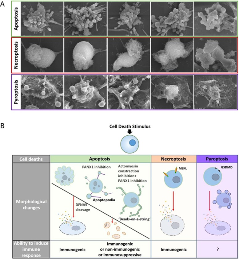
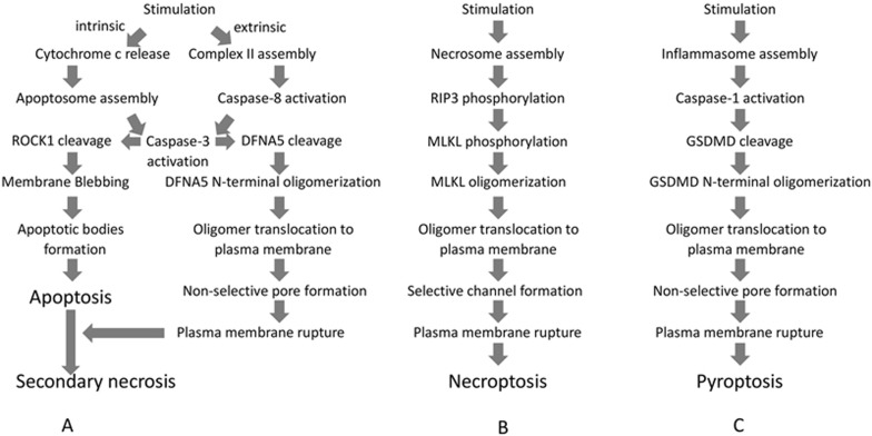
Ruptured and intact plasma membranes are classically considered as hallmarks of necrotic and apoptotic cell death, respectively. As such, apoptosis is usually considered a non-inflammatory process while necrosis triggers inflammation. Recent studies on necroptosis and pyroptosis, two types of programmed necrosis, revealed that plasma membrane rupture is mediated by MLKL channels during necroptosis but depends on non-selective gasdermin D (GSDMD) pores during pyroptosis. Importantly, the morphology of dying cells executed by MLKL channels can be distinguished from that executed by GSDMD pores. Interestingly, it was found recently that secondary necrosis of apoptotic cells, a previously believed non-regulated form of cell lysis that occurs after apoptosis, can be programmed and executed by plasma membrane pore formation like that of pyroptosis. In addition, pyroptosis is associated with pyroptotic bodies, which have some similarities to apoptotic bodies. Therefore, different cell death programs induce distinctive reshuffling processes of the plasma membrane. Given the fact that the nature of released intracellular contents plays a crucial role in dying/dead cell-induced immunogenicity, not only membrane rupture or integrity but also the nature of plasma membrane breakdown would determine the fate of a cell as well as its ability to elicit an immune response. In this review, we will discuss recent advances in the field of apoptosis, necroptosis and pyroptosis, with an emphasis on the mechanisms underlying plasma membrane changes observed on dying cells and their implication in cell death-elicited immunogenicity.
破裂和完整的质膜通常被认为分别是细胞坏死和细胞凋亡的标志。因此,凋亡通常被认为是非炎症过程,而坏死则引发炎症。最近关于细胞程序性坏死的两种类型——坏死性凋亡和细胞焦亡的研究表明,质膜破裂是由坏死性凋亡中的 MLKL 通道介导的,但依赖于细胞焦亡中的非选择性 gasdermin D(GSDMD)孔。重要的是,由 MLKL 通道介导的死亡细胞的形态可以与由 GSDMD 孔介导的形态区分开来。有趣的是,最近发现凋亡细胞的继发性坏死,即之前认为的凋亡后发生的非调控形式的细胞溶解,可以像细胞焦亡一样通过质膜孔形成进行编程和执行。此外,细胞焦亡与细胞焦亡小体有关,细胞焦亡小体与凋亡小体有一些相似之处。因此,不同的细胞死亡程序诱导质膜的不同重排过程。鉴于释放的细胞内物质的性质在死亡/坏死细胞诱导免疫原性中起着至关重要的作用,不仅膜破裂或完整性,而且质膜破裂的性质也将决定细胞的命运及其引发免疫反应的能力。在这篇综述中,我们将讨论细胞凋亡、坏死性凋亡和细胞焦亡领域的最新进展,重点讨论观察到的死亡细胞质膜变化的机制及其在细胞死亡引发的免疫原性中的意义。