• 文献检索
  • 文档翻译
  • 深度研究
  • 学术资讯
  • Suppr Zotero 插件Zotero 插件
  • 邀请有礼
  • 套餐&价格
  • 历史记录
应用&插件
Suppr Zotero 插件Zotero 插件浏览器插件Mac 客户端Windows 客户端微信小程序
定价
高级版会员购买积分包购买API积分包
服务
文献检索文档翻译深度研究API 文档MCP 服务
关于我们
关于 Suppr公司介绍联系我们用户协议隐私条款
关注我们

Suppr 超能文献

核心技术专利:CN118964589B侵权必究
粤ICP备2023148730 号-1Suppr @ 2026

文献检索

告别复杂PubMed语法,用中文像聊天一样搜索,搜遍4000万医学文献。AI智能推荐,让科研检索更轻松。

立即免费搜索

文件翻译

保留排版,准确专业,支持PDF/Word/PPT等文件格式,支持 12+语言互译。

免费翻译文档

深度研究

AI帮你快速写综述,25分钟生成高质量综述,智能提取关键信息,辅助科研写作。

立即免费体验

远视性屈光性角膜切削术后Pentacam HR与Orbscan II的比较。

Comparison between Pentacam HR and Orbscan II after Hyperopic Photorefractive Keratectomy.

作者信息

Jabbarvand Mahmoud, Askarizadeh Farshad, Sedaghat Mohamad Reza, Ghadimi Hadi, Khosravi Bahram, Amiri Mohammad Aghazadeh, Narooie-Noori Foroozan

机构信息

Eye Research Center, Farabi Eye Hospital, Tehran University of Medical Sciences, Tehran, Iran.

Department of Optometry, School of Paramedical Sciences, Mashhad University of Medical Sciences, Mashhad, Iran.

出版信息

J Ophthalmic Vis Res. 2017 Oct-Dec;12(4):374-379. doi: 10.4103/jovr.jovr_47_17.

DOI:10.4103/jovr.jovr_47_17
PMID:29090045
原文链接:https://pmc.ncbi.nlm.nih.gov/articles/PMC5644402/
Abstract

PURPOSE

The aim of this study was to determine the agreement between Pentacam HR (Scheimpflug imaging, Oculus) and Orbscan II (scanning slit topography, Bausch and Lomb) in measuring corneal parameters after photorefractive keratectomy (PRK) for hyperopia.

METHODS

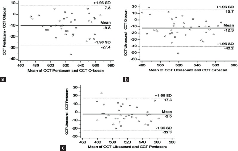
In this prospective cross-sectional study, 38 hyperopic eyes undergoing PRK were examined before refractive surgery and 8 to 10 months postoperatively using Pentacam HR and Orbscan II. Ultrasound (US) pachymetry was also used to measure central corneal thickness (CCT). The radius of anterior (A-) and posterior (P-) best-fit sphere size (BFS), central elevation (CE), and anterior maximum tangential power in 3 mm (TG3) and 3-5 mm (TG5) zones, anterior chamber depth (ACD), and central corneal thickness (CCT) were collected and used in the analyses. To study the agreement between the measurements made by the two devices, the method described by Bland and Altman was used and the 95% limits of agreement were calculated.

RESULTS

The 95% limits of agreement show reasonable agreement between the measurements by Pentacam HR and Orbscan II for A-BFS, P-BFS, A-TG3, and CCT, but not for A-CE, P-CE, A-TG5, or ACD. CCT values obtained by both Pentacam HR and Orbscan II correlated well with the values determined by US pachymetry.

CONCLUSION

Pentacam HR and Orbscan II after PRK for hyperopia show reasonable agreement for determining A-BFS, P-BFS, A-TG3, and CCT, but not for A-CE, P-CE, A-TG5, or ACD. CCT measurements with Pentacam HR have reasonable agreement with US pachymetry.

摘要

目的

本研究旨在确定Pentacam HR(眼前节分析系统,Oculus公司)与Orbscan II(扫描裂隙角膜地形图仪,博士伦公司)在测量远视性准分子激光原位角膜磨镶术(PRK)后角膜参数方面的一致性。

方法

在这项前瞻性横断面研究中,对38只接受PRK的远视眼在屈光手术前以及术后8至10个月使用Pentacam HR和Orbscan II进行检查。还使用超声(US)测厚法测量中央角膜厚度(CCT)。收集前(A-)和后(P-)最佳拟合球面尺寸(BFS)半径、中央高度(CE)、3mm(TG3)和3 - 5mm(TG5)区域的前表面最大切线屈光度、前房深度(ACD)以及中央角膜厚度(CCT)并用于分析。为研究两种设备测量结果之间的一致性,采用Bland和Altman描述的方法并计算95%一致性界限。

结果

95%一致性界限显示,Pentacam HR和Orbscan II在测量A - BFS、P - BFS、A - TG3和CCT方面具有合理的一致性,但在测量A - CE、P - CE、A - TG5或ACD方面则不然。Pentacam HR和Orbscan II获得的CCT值与US测厚法确定的值相关性良好。

结论

远视性PRK术后,Pentacam HR和Orbscan II在确定A - BFS、P - BFS、A - TG3和CCT方面显示出合理的一致性,但在A - CE、P - CE、A - TG5或ACD方面则不然。Pentacam HR测量的CCT与US测厚法具有合理的一致性。

https://cdn.ncbi.nlm.nih.gov/pmc/blobs/72f7/5644402/e6f3a21a8353/JOVR-12-374-g005.jpg
https://cdn.ncbi.nlm.nih.gov/pmc/blobs/72f7/5644402/6fd8f83aeaa5/JOVR-12-374-g003.jpg
https://cdn.ncbi.nlm.nih.gov/pmc/blobs/72f7/5644402/e6f3a21a8353/JOVR-12-374-g005.jpg
https://cdn.ncbi.nlm.nih.gov/pmc/blobs/72f7/5644402/6fd8f83aeaa5/JOVR-12-374-g003.jpg
https://cdn.ncbi.nlm.nih.gov/pmc/blobs/72f7/5644402/e6f3a21a8353/JOVR-12-374-g005.jpg

相似文献

1
Comparison between Pentacam HR and Orbscan II after Hyperopic Photorefractive Keratectomy.远视性屈光性角膜切削术后Pentacam HR与Orbscan II的比较。
J Ophthalmic Vis Res. 2017 Oct-Dec;12(4):374-379. doi: 10.4103/jovr.jovr_47_17.
2
Corneal changes after laser refractive surgery for myopia: comparison of Orbscan II and Pentacam findings.近视激光屈光手术后的角膜变化:Orbscan II与Pentacam检查结果比较
J Cataract Refract Surg. 2007 May;33(5):841-7. doi: 10.1016/j.jcrs.2007.01.019.
3
Central Corneal Thickness in Highly Myopic Eyes: Inter-device Agreement of Ultrasonic Pachymetry, Pentacam and Orbscan II Before and After Photorefractive Keratectomy.高度近视眼中的中央角膜厚度:准分子激光原位角膜磨镶术前后超声角膜测厚仪、Pentacam和Orbscan II的设备间一致性
J Ophthalmic Vis Res. 2014 Jan;9(1):14-21.
4
Pentacam and Orbscan II measurements of posterior corneal elevation before and after photorefractive keratectomy.准分子激光原位角膜磨镶术前后角膜后表面高度的Pentacam和Orbscan II测量
J Refract Surg. 2009 Mar;25(3):290-5. doi: 10.3928/1081597X-20090301-09.
5
Central corneal thickness measurement with Pentacam, Orbscan II, and ultrasound devices before and after laser refractive surgery for myopia.近视激光屈光手术后,使用Pentacam、Orbscan II和超声设备测量中央角膜厚度。
J Cataract Refract Surg. 2007 Oct;33(10):1701-7. doi: 10.1016/j.jcrs.2007.05.040.
6
Measurement of central corneal thickness using Orbscan 3, Pentacam HR and ultrasound pachymetry in normal eyes.正常眼的 Orbscan 3、Pentacam HR 和超声角膜测厚仪中央角膜厚度测量。
Int Ophthalmol. 2020 Jul;40(7):1759-1764. doi: 10.1007/s10792-020-01344-1. Epub 2020 Mar 27.
7
Repeatability and agreement of five imaging systems for measuring anterior segment parameters in healthy eyes.五种成像系统测量健康眼睛眼前节参数的重复性和一致性
Indian J Ophthalmol. 2017 Apr;65(4):288-294. doi: 10.4103/ijo.IJO_729_16.
8
Comparison of corneal measurements in keratoconic eyes using rotating Scheimpflug camera and scanning-slit topography.使用旋转式Scheimpflug相机和扫描裂隙地形图对圆锥角膜眼的角膜测量结果进行比较。
Int J Ophthalmol. 2015 Apr 18;8(2):275-80. doi: 10.3980/j.issn.2222-3959.2015.02.11. eCollection 2015.
9
Central corneal thickness measurements in unoperated eyes and eyes after PRK for myopia using Pentacam, Orbscan II, and ultrasonic pachymetry.使用Pentacam、Orbscan II和超声测厚法测量未手术眼和近视PRK术后眼的中央角膜厚度。
J Refract Surg. 2007 Nov;23(9):888-94. doi: 10.3928/1081-597X-20071101-04.
10
Central corneal thickness measurements after myopic photorefractive keratectomy using Scheimpflug imaging, scanning-slit topography, and ultrasonic pachymetry.使用 Scheimpflug 成像、扫描-slit topography 和超声角膜测厚仪测量近视性光折射性角膜切削术后的中央角膜厚度。
J Cataract Refract Surg. 2010 Sep;36(9):1543-9. doi: 10.1016/j.jcrs.2010.03.042.

引用本文的文献

1
Comparison of central corneal thickness in healthy eyes using ultrasound pachymetry, non-contact specular microscopy and a high-resolution Scheimpflug camera.使用超声测厚法、非接触式角膜内皮显微镜和高分辨率Scheimpflug相机对健康眼睛的中央角膜厚度进行比较。
Int Ophthalmol. 2023 Feb;43(2):363-370. doi: 10.1007/s10792-022-02431-1. Epub 2022 Jul 21.
2
Author's Reply.作者回复。
J Ophthalmic Vis Res. 2018 Jul-Sep;13(3):366-367. doi: 10.4103/jovr.jovr_97_18.
3
Corneal Thickness Measurement after Hyperopic Photorefractive Keratectomy.

本文引用的文献

1
Corneal Regeneration After Photorefractive Keratectomy: A Review.准分子激光原位角膜磨镶术后的角膜再生:综述
J Optom. 2015 Jul-Sep;8(3):149-69. doi: 10.1016/j.optom.2014.09.001. Epub 2014 Oct 23.
2
Repeatability, reproducibility, and agreement between three different Scheimpflug systems in measuring corneal and anterior segment biometry.三种不同 Scheimpflug 系统测量角膜和眼前节生物测量的重复性、可再现性和一致性。
J Refract Surg. 2014 Sep;30(9):616-21. doi: 10.3928/1081597X-20140815-02.
3
Measurements of anterior segment parameters using three different non-contact optical devices in keratoconus patients.
远视性屈光性角膜切削术后角膜厚度测量
J Ophthalmic Vis Res. 2018 Jul-Sep;13(3):365. doi: 10.4103/jovr.jovr_217_17.
在圆锥角膜患者中使用三种不同的非接触式光学设备测量眼前节参数。
Int J Ophthalmol. 2013 Aug 18;6(4):521-5. doi: 10.3980/j.issn.2222-3959.2013.04.21. eCollection 2013.
4
Comparison and repeatability of keratometric and corneal power measurements obtained by Orbscan II, Pentacam, and Galilei corneal tomography systems.眼前节分析系统 Orbscan II、Pentacam 和 Galilei 角膜地形图仪测量的角膜曲率和角膜屈光度的比较及可重复性。
Am J Ophthalmol. 2013 Jul;156(1):53-60. doi: 10.1016/j.ajo.2013.01.029. Epub 2013 Mar 28.
5
Corneal thickness measurement using Orbscan, Pentacam, Galilei, and ultrasound in normal and post-femtosecond laser in situ keratomileusis eyes.应用 Orbscan、Pentacam、Galilei 和超声角膜测厚仪测量正常眼和飞秒激光原位角膜磨镶术后眼的角膜厚度。
Cornea. 2012 Sep;31(9):978-82. doi: 10.1097/ICO.0b013e31823d03fc.
6
Imaging of the cornea: topography vs tomography.角膜成像:地形图与断层扫描
J Refract Surg. 2010 Nov;26(11):847-9. doi: 10.3928/1081597X-20101006-01.
7
Corneal imaging with slit-scanning and Scheimpflug imaging techniques.
Clin Exp Optom. 2011 Jan;94(1):33-42. doi: 10.1111/j.1444-0938.2010.00509.x. Epub 2010 Aug 16.
8
Central corneal thickness measurements after myopic photorefractive keratectomy using Scheimpflug imaging, scanning-slit topography, and ultrasonic pachymetry.使用 Scheimpflug 成像、扫描-slit topography 和超声角膜测厚仪测量近视性光折射性角膜切削术后的中央角膜厚度。
J Cataract Refract Surg. 2010 Sep;36(9):1543-9. doi: 10.1016/j.jcrs.2010.03.042.
9
Comparison of corneal thickness measurements using Galilei, HR Pentacam, and ultrasound.使用 Galilei、HR Pentacam 和超声测量角膜厚度的比较。
Cornea. 2010 Oct;29(10):1091-5. doi: 10.1097/ICO.0b013e3181cf98e5.
10
Intraocular lens power calculation after corneal refractive surgery.角膜屈光手术后的人工晶状体度数计算。
Middle East Afr J Ophthalmol. 2010 Jan;17(1):63-8. doi: 10.4103/0974-9233.61219.