Pamon Tee, Bhandal Vincent, Adler Benjamin J, Ete Chan M, Rubin Clinton T
Department of Biomedical Engineering, Stony Brook University, Stony Brook, NY, 11794-5281.
J Orthop Res. 2018 Feb;36(2):751-759. doi: 10.1002/jor.23795. Epub 2017 Dec 5.
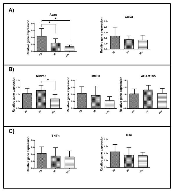
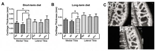
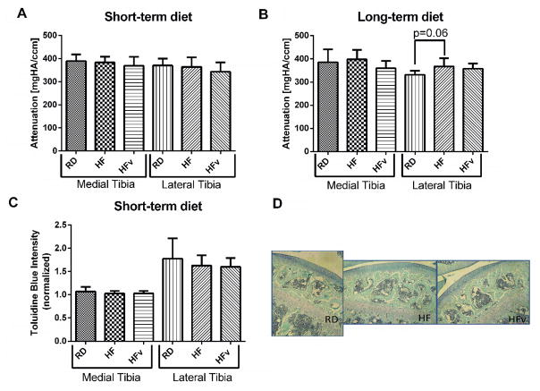
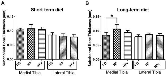
Obesity is associated with an elevated risk of osteoarthritis (OA). We examined here whether high fat diet administered in young mice, compromised the attainment of articular cartilage thickness. Further, we sought to determine if low-intensity vibration (LIV) could protect the retention of articular cartilage in a mouse model of diet-induced obesity. Five-week-old, male, C57BL/6 mice were separated into three groups (n = 10): Regular diet (RD), High fat diet (HF), and HF + LIV (HFv; 90 Hz, 0.2g, 30 min/d, 5 d/w) administered for 6 weeks. Additionally, an extended HF diet study was run for 6 months (LIV at 15 m/d). Articular cartilage and subchondral bone morphology, and sulfated GAG content were quantified using contrast agent enhanced μCT and histology. Gene expression within femoral condyles was quantified using real-time polymerase chain reaction. Contrary to our hypothesis, HF cartilage thickness was not statistically different from RD. However, LIV increased cartilage thickness compared to HF, and the elevated thickness was maintained when diet and LIV were extended into adulthood. RT-PCR analysis showed a reduction of aggrecan expression with high fat diet, while application of LIV reduced the expression of degradative MMP-13. Further, long-term HF diet resulted in subchondral bone thickening, compared to RD, providing early evidence of OA pathology-LIV suppressed the thickening, such that levels were not significantly different from RD. These data suggest that dynamic loading, via LIV, protected the retention of cartilage thickness, potentially resulting in joint surfaces better suited to endure the risks of elevated loading that parallel obesity. © 2017 Orthopaedic Research Society. Published by Wiley Periodicals, Inc. J Orthop Res 36:751-759, 2018.
肥胖与骨关节炎(OA)风险升高相关。我们在此研究了给年轻小鼠喂食高脂肪饮食是否会影响关节软骨厚度的增长。此外,我们试图确定低强度振动(LIV)能否在饮食诱导的肥胖小鼠模型中保护关节软骨。将5周龄的雄性C57BL/6小鼠分为三组(n = 10):常规饮食(RD)组、高脂肪饮食(HF)组和高脂肪饮食+低强度振动(HFv;90Hz,0.2g,每天30分钟,每周5天)组,持续喂食6周。另外,进行了一项为期6个月的延长高脂肪饮食研究(每天15米的低强度振动)。使用造影剂增强微计算机断层扫描(μCT)和组织学方法对关节软骨和软骨下骨形态以及硫酸化糖胺聚糖(GAG)含量进行定量分析。使用实时聚合酶链反应对股骨髁内的基因表达进行定量分析。与我们的假设相反,高脂肪饮食组的软骨厚度与常规饮食组相比无统计学差异。然而,与高脂肪饮食组相比,低强度振动增加了软骨厚度,并且当饮食和低强度振动延长至成年期时,增加的厚度得以维持。逆转录-聚合酶链反应(RT-PCR)分析表明,高脂肪饮食会使聚集蛋白聚糖表达降低,而低强度振动的应用则降低了降解性基质金属蛋白酶-13(MMP-13)的表达。此外,与常规饮食组相比,长期高脂肪饮食导致软骨下骨增厚,这为骨关节炎病理提供了早期证据——低强度振动抑制了增厚,使其水平与常规饮食组无显著差异。这些数据表明,通过低强度振动进行的动态加载保护了软骨厚度,可能使关节表面更适合承受与肥胖相关的负荷增加风险。©2017骨科研究协会。由威利期刊公司出版。《骨科研究杂志》36:751 - 759,2018年。