Division of RI-Convergence Research, Korea Institute of Radiological & Medical Sciences, Seoul, Republic of Korea.
Convergence Institute of Biomedical Engineering and Biomaterials, Seoul National University of Science & Technology, Seoul, Republic of Korea.
Contrast Media Mol Imaging. 2017 Sep 26;2017:3981358. doi: 10.1155/2017/3981358. eCollection 2017.
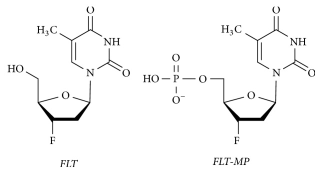
The thymidine analogue 3'-deoxy-3'-[F]fluorothymidine, or [F]fluorothymidine ([F]FLT), is used to measure tumor cell proliferation with positron emission tomography (PET) imaging technology in nuclear medicine. FLT is phosphorylated by thymidine kinase 1 (TK1) and then trapped inside cells; it is not incorporated into DNA. Imaging with F-radiolabeled FLT is a noninvasive technique to visualize cellular proliferation in tumors. However, it is difficult to distinguish between [F]FLT and its metabolites by PET imaging, and quantification has not been attempted using current imaging methods. In this study, we successfully acquired F spectra of natural or nonradioactive 3'-deoxy-3'-fluorothymidine ([F]FLT) and its monophosphate metabolite (FLT-MP) in a tumor xenograft mouse model using 9.4T magnetic resonance imaging (MRI). This preliminary result demonstrates that F magnetic resonance spectroscopy (MRS) with FLT is suitable for the assessment of tumor aggressiveness and for early prediction of treatment response.
胸苷类似物 3'-脱氧-3'-[F]氟代胸苷,或[F]氟代胸苷([F]FLT),用于通过核医学中的正电子发射断层扫描 (PET) 成像技术测量肿瘤细胞增殖。FLT 被胸苷激酶 1 (TK1) 磷酸化,然后被捕获在细胞内;它不被掺入 DNA 中。用 F 放射性标记的 FLT 成像是非侵入性技术,可在肿瘤中可视化细胞增殖。然而,通过 PET 成像很难区分[F]FLT 和其代谢物,并且当前的成像方法尚未尝试定量。在这项研究中,我们使用 9.4T 磁共振成像 (MRI) 在肿瘤异种移植小鼠模型中成功地获得了天然或非放射性 3'-脱氧-3'-氟代胸苷([F]FLT)及其单磷酸代谢物(FLT-MP)的 F 谱。这个初步结果表明,FLT 的 F 磁共振波谱 (MRS) 适合评估肿瘤侵袭性,并可早期预测治疗反应。