Hurrell Sarah L, McGarry Sean D, Kaczmarowski Amy, Iczkowski Kenneth A, Jacobsohn Kenneth, Hohenwalter Mark D, Hall William A, See William A, Banerjee Anjishnu, Charles David K, Nevalainen Marja T, Mackinnon Alexander C, LaViolette Peter S
Medical College of Wisconsin, Department of Radiology, Milwaukee, Wisconsin, United States.
Medical College of Wisconsin, Department of Biophysics, Milwaukee, Wisconsin, United States.
J Med Imaging (Bellingham). 2018 Jan;5(1):011004. doi: 10.1117/1.JMI.5.1.011004. Epub 2017 Oct 27.
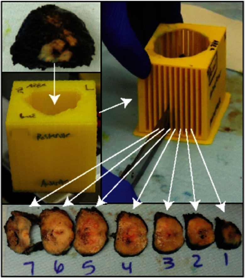
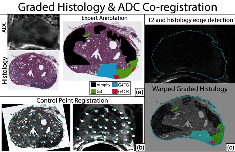
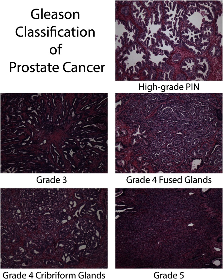
Multiparametric magnetic resonance imaging (MP-MRI), including diffusion-weighted imaging, is commonly used to diagnose prostate cancer. This radiology-pathology study correlates prostate cancer grade and morphology with common -value combinations for calculating apparent diffusion coefficient (ADC). Thirty-nine patients undergoing radical prostatectomy were recruited for MP-MRI prior to surgery. Diffusion imaging was collected with seven -values, and ADC was calculated. Excised prostates were sliced in the same orientation as the MRI using 3-D printed slicing jigs. Whole-mount slides were digitized and annotated by a pathologist. Annotated samples were aligned to the MRI, and ADC values were extracted from annotated peripheral zone (PZ) regions. A receiver operating characteristic (ROC) analysis was performed to determine accuracy of tissue type discrimination and optimal ADC -value combination. ADC significantly discriminates Gleason (G) G4-5 cancer from G3 and other prostate tissue types. The optimal -values for discriminating high from low-grade and noncancerous tissue in the PZ are 50 and 2000, followed closely by 100 to 2000 and 0 to 2000. Optimal ADC cut-offs are presented for dichotomized discrimination of tissue types according to each -value combination. Selection of -values affects the sensitivity and specificity of ADC for discrimination of prostate cancer.
多参数磁共振成像(MP-MRI),包括扩散加权成像,常用于诊断前列腺癌。这项放射学-病理学研究将前列腺癌的分级和形态与计算表观扩散系数(ADC)的常见值组合相关联。39例行根治性前列腺切除术的患者在手术前接受了MP-MRI检查。采用7个值进行扩散成像,并计算ADC。使用3D打印切片夹具将切除的前列腺切成与MRI相同的方向。病理学家对整装切片进行数字化处理并标注。将标注后的样本与MRI对齐,并从标注的外周带(PZ)区域提取ADC值。进行了受试者操作特征(ROC)分析,以确定组织类型鉴别和最佳ADC值组合的准确性。ADC能显著区分Gleason(G)4-5级癌与G3级癌及其他前列腺组织类型。在PZ中区分高级别与低级别及非癌组织的最佳值分别为50和2000,紧随其后的是100至2000和0至2000。根据每个值组合给出了用于组织类型二分法鉴别的最佳ADC临界值。值的选择会影响ADC鉴别前列腺癌的敏感性和特异性。