Fortes Marco A S, Scervino Maria V M, Marzuca-Nassr Gabriel N, Vitzel Kaio F, da Justa Pinheiro Carlos H, Curi Rui
Department of Physiology and Biophysics, Institute of Biomedical Sciences, University of São Paulo, São Paulo, Brazil.
Department of Internal Medicine, Faculty of Medicine, Universidad de La Frontera, Temuco, Chile.
Front Physiol. 2017 Oct 26;8:830. doi: 10.3389/fphys.2017.00830. eCollection 2017.
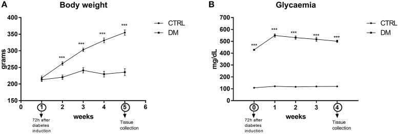
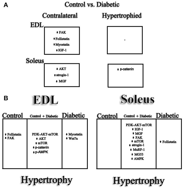
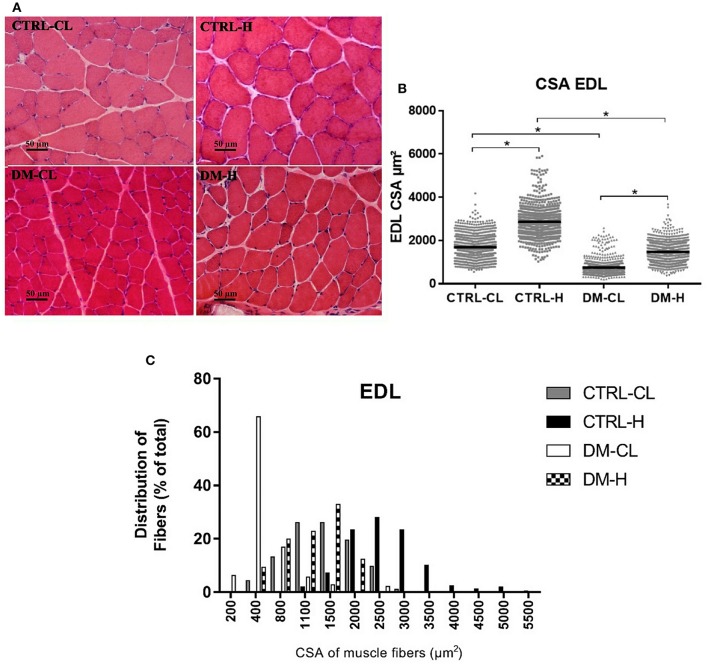
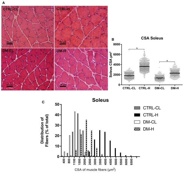
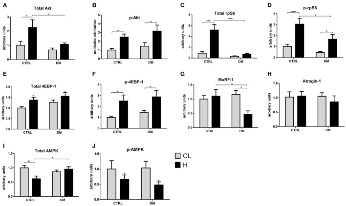
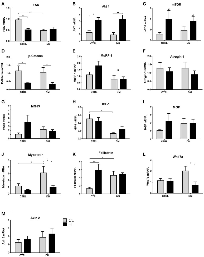
Diabetes mellitus induces a reduction in skeletal muscle mass and strength. Strength training is prescribed as part of treatment since it improves glycemic control and promotes increase of skeletal muscle mass. The mechanisms involved in overload-induced muscle hypertrophy elicited at the establishment of the type I diabetic state was investigated in Wistar rats. The purpose was to examine whether the overload-induced hypertrophy can counteract the hypotrophy associated to the diabetic state. The experiments were performed in oxidative (soleus) or glycolytic (EDL) muscles. PI3K/Akt/mTOR protein synthesis pathway was evaluated 7 days after overload-induced hypertrophy of soleus and of EDL muscles. The mRNA expression of genes associated with different signaling pathways that control muscle hypertrophy was also evaluated: mechanotransduction (FAK), Wnt/β-catenin, myostatin, and follistatin. The soleus and EDL muscles when submitted to overload had similar hypertrophic responses in control and diabetic animals. The increase of absolute and specific twitch and tetanic forces had the same magnitude as muscle hypertrophic response. Hypertrophy of the EDL muscle from diabetic animals mostly involved mechanical loading-stimulated PI3K/Akt/mTOR pathway besides the reduced activation of AMP-activated protein kinase (AMPK) and decrease of myostatin expression. Hypertrophy was more pronounced in the soleus muscle of diabetic animals due to a more potent activation of rpS6 and increased mRNA expression of insulin-like growth factor-1 (IGF-1), mechano-growth factor (MGF) and follistatin, and decrease of myostatin, MuRF-1 and atrogin-1 contents. The signaling changes enabled the soleus muscle mass and force of the diabetic rats to reach the values of the control group.
糖尿病会导致骨骼肌质量和力量下降。力量训练作为治疗的一部分被推荐,因为它能改善血糖控制并促进骨骼肌质量增加。在Wistar大鼠中研究了I型糖尿病状态建立时由超负荷引起的肌肉肥大所涉及的机制。目的是检查超负荷诱导的肥大是否能抵消与糖尿病状态相关的萎缩。实验在氧化型(比目鱼肌)或糖酵解型(趾长伸肌)肌肉中进行。在比目鱼肌和趾长伸肌超负荷诱导肥大7天后,评估PI3K/Akt/mTOR蛋白合成途径。还评估了与控制肌肉肥大的不同信号通路相关基因的mRNA表达:机械转导(黏着斑激酶)、Wnt/β-连环蛋白、肌肉生长抑制素和卵泡抑素。在对照动物和糖尿病动物中,比目鱼肌和趾长伸肌在承受超负荷时具有相似的肥大反应。绝对和特定的抽搐及强直力量的增加与肌肉肥大反应幅度相同。糖尿病动物趾长伸肌的肥大除了涉及机械负荷刺激的PI3K/Akt/mTOR途径外,还包括AMP激活的蛋白激酶(AMPK)激活减少和肌肉生长抑制素表达降低。由于rpS6的更强激活以及胰岛素样生长因子-1(IGF-1)、机械生长因子(MGF)和卵泡抑素的mRNA表达增加,以及肌肉生长抑制素、肌肉泛素连接酶-1(MuRF-1)和肌肉萎缩相关基因-1(atrogin-1)含量降低,糖尿病动物比目鱼肌的肥大更为明显。这些信号变化使糖尿病大鼠的比目鱼肌质量和力量达到对照组的值。