Aggarwal Kanika, Agarwal Aniruddha, Katoch Deeksha, Sharma Mansi, Gupta Vishali
Advanced Eye Center, Postgraduate Institute of Medical Education and Research, Chandigarh, India.
Indian J Ophthalmol. 2017 Nov;65(11):1235-1238. doi: 10.4103/ijo.IJO_485_17.
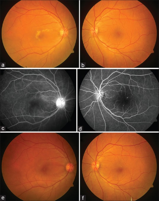
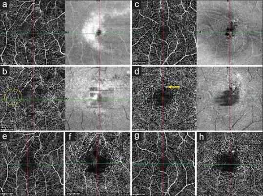
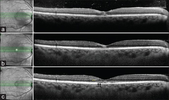
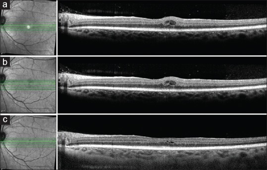
A 39-year-old female with dengue fever presented with decreased vision in both eyes. Visual acuity was 20/200 and 20/80 in the right eye (OD) and left eye (OS), respectively. Fundus showed granular, grayish-white lesions in the parafoveal region in OD. Multimodal imaging including optical coherence tomography angiography (OCTA), optical coherence tomography (OCT), and fluorescein angiography (FA) was performed. FA showed late hyperfluorescence with few microaneurysms in OS. OCT showed hyperreflectivity in various layers, suggestive of acute macular neuroretinopathy (AMN). OCTA showed disruption of retinal capillary plexuses. This case shows how OCTA provides newer insights into the pathogenesis of AMN lesions in dengue fever.
一名39岁的登革热女性患者出现双眼视力下降。右眼(OD)和左眼(OS)的视力分别为20/200和20/80。眼底检查显示OD的黄斑旁区域有颗粒状灰白色病变。进行了包括光学相干断层扫描血管造影(OCTA)、光学相干断层扫描(OCT)和荧光素血管造影(FA)在内的多模态成像检查。FA显示OS有晚期高荧光,伴有少量微动脉瘤。OCT显示各层均有高反射性,提示急性黄斑神经视网膜病变(AMN)。OCTA显示视网膜毛细血管丛中断。该病例展示了OCTA如何为登革热中AMN病变的发病机制提供新的见解。