• 文献检索
  • 文档翻译
  • 深度研究
  • 学术资讯
  • Suppr Zotero 插件Zotero 插件
  • 邀请有礼
  • 套餐&价格
  • 历史记录
应用&插件
Suppr Zotero 插件Zotero 插件浏览器插件Mac 客户端Windows 客户端微信小程序
定价
高级版会员购买积分包购买API积分包
服务
文献检索文档翻译深度研究API 文档MCP 服务
关于我们
关于 Suppr公司介绍联系我们用户协议隐私条款
关注我们

Suppr 超能文献

核心技术专利:CN118964589B侵权必究
粤ICP备2023148730 号-1Suppr @ 2026

文献检索

告别复杂PubMed语法,用中文像聊天一样搜索,搜遍4000万医学文献。AI智能推荐,让科研检索更轻松。

立即免费搜索

文件翻译

保留排版,准确专业,支持PDF/Word/PPT等文件格式,支持 12+语言互译。

免费翻译文档

深度研究

AI帮你快速写综述,25分钟生成高质量综述,智能提取关键信息,辅助科研写作。

立即免费体验

不同年龄组健康志愿者的角膜清晰度测量:观察性研究。

Corneal clarity measurements in healthy volunteers across different age groups: Observational study.

作者信息

Alzahrani Khaled, Carley Fiona, Brahma Arun, Morley Debbie, Hillarby M Chantal

机构信息

aDivision of Pharmacy and Optometry, School of Health Sciences, University of Manchester bManchester Royal Eye Hospital, Manchester, UK.

出版信息

Medicine (Baltimore). 2017 Nov;96(46):e8563. doi: 10.1097/MD.0000000000008563.

DOI:10.1097/MD.0000000000008563
PMID:29145265
原文链接:https://pmc.ncbi.nlm.nih.gov/articles/PMC5704810/
Abstract

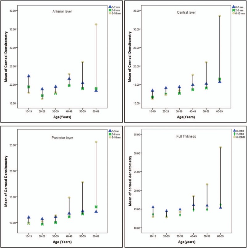
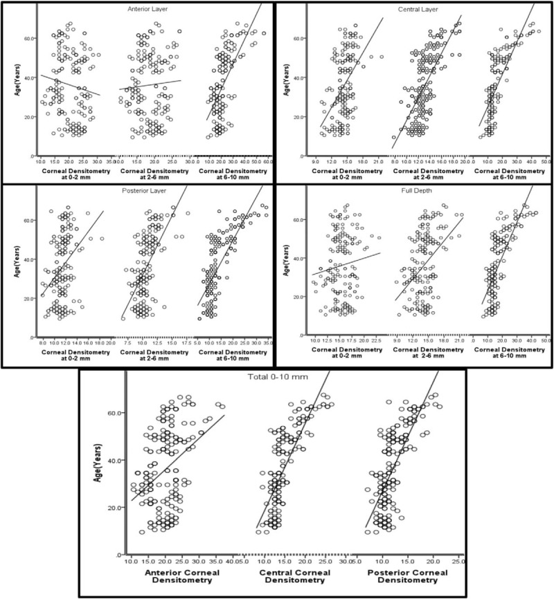
The aim of this study was to standardize and investigate the changes in corneal clarity with age. Densitometry software for the Oculus Pentacam was used to examine corneal clarity at different age groups.A total of 192 eyes from 97 healthy participants were included in this cohort comparative nonrandomized, cross-sectional study. An Oculus Pentcam was used to image the cornea of healthy participants grouped by age (between 10 and 70 years old). Data from the densitometry output have been used to determine clarity in concentric zones and different depths of the cornea.Corneal densitometry (CD) across all ages showed significant differences between groups when divided into the following layers: anterior, central, and posterior or divided into 0 to 2, 2 to 6, and 6 to 10 mm concentric zones (P < .05). The most striking decrease in clarity occurred with age in all 3 layers of the periphery (6-10 mm) (P < .05). In addition, we showed that the 10 to 19-year age group had lower clarity than the 20 to 30-age group (P < .05), and after 30 years, the cornea shows a steady progression of increased or decreased clarity.The values for CD, as well as for separate subdivisions based on layer and surface area, might provide a standard for use in further studies and clinical practice. This study established that relation between CD and age is differed when the cornea is divided into layers and zones. This study suggests that there are other factors that may play an essential role in corneal clarity as well as age.

摘要

本研究的目的是规范并调查角膜透明度随年龄的变化。使用Oculus Pentacam的密度测定软件检查不同年龄组的角膜透明度。本队列比较性非随机横断面研究纳入了97名健康参与者的192只眼睛。使用Oculus Pentcam对按年龄分组(10至70岁)的健康参与者的角膜进行成像。密度测定输出的数据已用于确定角膜同心区域和不同深度的透明度。当按以下层划分:前部、中央和后部,或划分为0至2、2至6和6至10毫米的同心区域时,各年龄组之间的角膜密度测定(CD)显示出显著差异(P<0.05)。周边所有三层(6至10毫米)的透明度随年龄增长下降最为显著(P<0.05)。此外,我们发现10至19岁年龄组的透明度低于20至30岁年龄组(P<0.05),30岁以后,角膜透明度呈现出增加或减少的稳定变化趋势。CD值以及基于层和表面积的单独细分值可能为进一步研究和临床实践提供一个标准。本研究确定,当将角膜分为层和区域时,CD与年龄之间的关系有所不同。本研究表明,除年龄外,可能还有其他因素在角膜透明度中起重要作用。

https://cdn.ncbi.nlm.nih.gov/pmc/blobs/542e/5704810/b38392724823/medi-96-e8563-g005.jpg
https://cdn.ncbi.nlm.nih.gov/pmc/blobs/542e/5704810/a23a6f6a5ba8/medi-96-e8563-g003.jpg
https://cdn.ncbi.nlm.nih.gov/pmc/blobs/542e/5704810/b38392724823/medi-96-e8563-g005.jpg
https://cdn.ncbi.nlm.nih.gov/pmc/blobs/542e/5704810/a23a6f6a5ba8/medi-96-e8563-g003.jpg
https://cdn.ncbi.nlm.nih.gov/pmc/blobs/542e/5704810/b38392724823/medi-96-e8563-g005.jpg

相似文献

1
Corneal clarity measurements in healthy volunteers across different age groups: Observational study.不同年龄组健康志愿者的角膜清晰度测量:观察性研究。
Medicine (Baltimore). 2017 Nov;96(46):e8563. doi: 10.1097/MD.0000000000008563.
2
Normative values for corneal densitometry analysis by Scheimpflug optical assessment.Scheimpflug 光学评估角膜密度分析的正常值。
Invest Ophthalmol Vis Sci. 2014 Jan 7;55(1):162-8. doi: 10.1167/iovs.13-13236.
3
Corneal densitometry and its correlation with age, pachymetry, corneal curvature, and refraction.角膜密度测定及其与年龄、角膜厚度测量、角膜曲率和屈光的相关性。
Int Ophthalmol. 2017 Dec;37(6):1263-1268. doi: 10.1007/s10792-016-0397-y. Epub 2016 Nov 11.
4
Quantitative assessment of corneal clarity in keratoconus: a case control study of corneal densitometry.圆锥角膜中角膜透明度的定量评估:角膜密度测定的病例对照研究
Eur J Ophthalmol. 2016 Jan-Feb;26(1):18-23. doi: 10.5301/ejo.5000644. Epub 2015 Jul 7.
5
Corneal Densitometry: A New Technique for Objective Assessment of Corneal Clarity in Pseudoexfoliation Syndrome.角膜密度测定法:一种用于客观评估假性剥脱综合征角膜透明度的新技术。
J Glaucoma. 2016 Sep;25(9):775-9. doi: 10.1097/IJG.0000000000000501.
6
Assessment of corneal backward light scattering in the healthy cornea and factors affecting corneal transparency.健康角膜中角膜向后光散射的评估及影响角膜透明度的因素。
Jpn J Ophthalmol. 2018 May;62(3):335-341. doi: 10.1007/s10384-018-0584-7. Epub 2018 Mar 16.
7
The effect of topical latanoprost on corneal clarity; 1-year prospective study.局部用拉坦前列素对角膜透明度的影响:1年前瞻性研究。
Cutan Ocul Toxicol. 2019 Sep;38(3):253-257. doi: 10.1080/15569527.2019.1590390. Epub 2019 Mar 25.
8
Evaluation of corneal and lens clarity in unilateral pseudoexfoliation syndrome: a densitometric analysis.单侧假性剥脱综合征中角膜和晶状体透明度的评估:密度测定分析
Clin Exp Optom. 2018 Nov;101(6):740-746. doi: 10.1111/cxo.12802. Epub 2018 Jun 21.
9
Objective Evaluation of Corneal and Lens Clarity in Children With Type 1 Diabetes Mellitus.1型糖尿病患儿角膜和晶状体清晰度的客观评估
Am J Ophthalmol. 2017 Jul;179:190-197. doi: 10.1016/j.ajo.2017.05.010. Epub 2017 May 17.
10
Age-Related Corneal Transparency Changes Evaluated With an Alternative Method to Corneal Densitometry.用角膜密度计替代方法评估年龄相关的角膜透明度变化。
Cornea. 2021 Feb 1;40(2):215-222. doi: 10.1097/ICO.0000000000002511.

引用本文的文献

1
Corneal densitometry in Chinese adults with healthy corneas: associations with sex, age, ocular metrics, and optical characteristics.健康角膜的中国成年人角膜密度测定:与性别、年龄、眼参数和光学特征的关系。
BMC Ophthalmol. 2024 May 31;24(1):230. doi: 10.1186/s12886-024-03500-y.
2
The influence of pterygium on corneal densitometry evaluated using the Oculus Pentacam system.翼状胬肉对使用欧科路Pentacam系统评估的角膜密度测定的影响。
Front Med (Lausanne). 2023 Jun 15;10:1184318. doi: 10.3389/fmed.2023.1184318. eCollection 2023.
3
Porcine Cornea Storage Ex Vivo Model as an Alternative to Human Donor Tissues for Investigations of Endothelial Layer Preservation.

本文引用的文献

1
Wound healing process after corneal stromal thinning observed with anterior segment optical coherence tomography.眼前节光学相干断层扫描观察角膜基质变薄后的伤口愈合过程。
Cornea. 2014 Oct;33(10):1056-60. doi: 10.1097/ICO.0000000000000223.
2
Long-term outcomes of boston type 1 keratoprosthesis implantation: a retrospective multicenter cohort.Boston 1 型角膜移植术的长期疗效:回顾性多中心队列研究。
Ophthalmology. 2014 Nov;121(11):2159-64. doi: 10.1016/j.ophtha.2014.05.030. Epub 2014 Jul 11.
3
Corneal epithelial remodeling following cataract surgery: three-dimensional investigation with anterior-segment optical coherence tomography.
猪角膜离体储存模型作为研究内皮细胞层保存的人供体组织替代物。
Transl Vis Sci Technol. 2023 Apr 3;12(4):24. doi: 10.1167/tvst.12.4.24.
4
Age-related changes in Scheimpflug corneal densitometry and their correlations with corneal topographic measurements in a healthy Chinese population.中国健康人群中,基于Scheimpflug角膜密度测量法的年龄相关性变化及其与角膜地形图测量结果的相关性
Quant Imaging Med Surg. 2023 Mar 1;13(3):1631-1641. doi: 10.21037/qims-22-486. Epub 2023 Feb 3.
5
Long-term changes in corneal densitometry and associated factors following small incision lenticule extraction for moderate and high myopia.中高度近视小切口透镜切除术术后角膜密度测量的长期变化及相关因素
Front Med (Lausanne). 2022 Sep 8;9:945894. doi: 10.3389/fmed.2022.945894. eCollection 2022.
6
Influence of eye tilt on corneal densitometry.眼倾斜对角膜密度测定的影响。
Ophthalmic Physiol Opt. 2022 Sep;42(5):1032-1037. doi: 10.1111/opo.13020. Epub 2022 Jun 16.
7
Corneal Imaging and Densitometry Measurements in Juvenile and Adult Keratoconus Patients to Evaluate Disease Progression and Treatment Effects After Corneal Cross-Linking.青少年和成人圆锥角膜患者的角膜成像和密度测量,以评估角膜交联术后的疾病进展和治疗效果。
Clin Optom (Auckl). 2019 Dec 30;11:173-180. doi: 10.2147/OPTO.S226000. eCollection 2019.
8
Influence of peripheral corneal relaxing incisions during cataract surgery for corneal astigmatism up to 2.5 dioptres on corneal densitometry.白内障手术中 2.5 屈光度以下周边角膜松解切口对角膜密度测量的影响。
Eye (Lond). 2019 May;33(5):804-811. doi: 10.1038/s41433-018-0327-5. Epub 2019 Jan 2.
9
Quantitative evaluation of corneal epithelial edema after cataract surgery using corneal densitometry: a prospective study.使用角膜密度计对白内障手术后角膜上皮水肿进行定量评估:一项前瞻性研究。
BMC Ophthalmol. 2018 Dec 20;18(1):334. doi: 10.1186/s12886-018-0998-5.
10
Corneal imaging and densitometry measurements in keratoconus patients to monitor disease progression and treatment outcomes after contact lens or Intacs treatment.圆锥角膜患者的角膜成像和密度测量,以监测隐形眼镜或Intacs治疗后的疾病进展和治疗效果。
Clin Ophthalmol. 2018 Sep 4;12:1653-1658. doi: 10.2147/OPTH.S168865. eCollection 2018.
白内障手术后角膜上皮重塑:前节光学相干断层扫描的三维研究。
J Refract Surg. 2014 May;30(5):348-53. doi: 10.3928/1081597X-20140416-04.
4
Epithelial remodeling as basis for machine-based identification of keratoconus.基于上皮重塑的角膜膨隆机识别。
Invest Ophthalmol Vis Sci. 2014 Mar 13;55(3):1580-7. doi: 10.1167/iovs.13-12578.
5
Normative values for corneal densitometry analysis by Scheimpflug optical assessment.Scheimpflug 光学评估角膜密度分析的正常值。
Invest Ophthalmol Vis Sci. 2014 Jan 7;55(1):162-8. doi: 10.1167/iovs.13-13236.
6
SD-OCT analysis of regional epithelial thickness profiles in keratoconus, postoperative corneal ectasia, and normal eyes.SD-OCT 分析圆锥角膜、术后角膜扩张和正常眼中的区域性上皮厚度曲线。
J Refract Surg. 2013 Mar;29(3):173-9. doi: 10.3928/1081597X-20130129-08.
7
Corneal white-to-white distance and mesopic pupil diameter.角膜白对白距离及中间视觉瞳孔直径。
Int J Ophthalmol. 2012;5(4):505-9. doi: 10.3980/j.issn.2222-3959.2012.04.19. Epub 2012 Aug 18.
8
Significant visual increase following infectious keratitis after collagen cross-linking.感染性角膜炎行胶原交联术后视力显著提高。
J Refract Surg. 2012 Aug;28(8):587-8. doi: 10.3928/1081597X-20120628-01. Epub 2012 Jul 13.
9
Normative database for corneal backscatter analysis by in vivo confocal microscopy.基于活体共聚焦显微镜的角膜背散射分析的规范数据库。
Invest Ophthalmol Vis Sci. 2011 Sep 21;52(10):7274-81. doi: 10.1167/iovs.11-7747.
10
Computerized Scheimpflug densitometry as a measure of corneal optical density after excimer laser refractive surgery in myopic eyes.计算机化 Scheimpflug 密度测定术作为近视眼角膜屈光手术后角膜光学密度的测量方法。
J Cataract Refract Surg. 2011 Aug;37(8):1502-6. doi: 10.1016/j.jcrs.2011.03.037.