Lawson Health Research Institute, St Joseph's Health Care, 268 Grosvenor St., London, Ontario N6A 4V2, Canada; Department of Medical Biophysics, Schulich School of Medicine and Dentistry, Western University, Medical Sciences Building, Rm M407, London, Ontario N6A 5C1, Canada.
Department of Clinical Neurological Sciences, Western University, 339 Windermere Road, London, Ontario N6A 5A5, Canada.
Neuroimage Clin. 2017 Oct 31;17:405-414. doi: 10.1016/j.nicl.2017.10.033. eCollection 2018.
The clinical utility of FDG-PET in diagnosing frontotemporal dementia (FTD) has been well demonstrated over the past decades. On the contrary, the diagnostic value of arterial spin labelling (ASL) MRI - a relatively new technique - in clinical diagnosis of FTD has yet to be confirmed. Using simultaneous PET/MRI, we evaluated the diagnostic performance of ASL in identifying pathological abnormalities in FTD (FTD) to determine whether ASL can provide similar diagnostic value as FDG-PET.
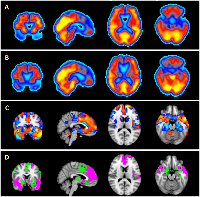
ASL and FDG-PET images were compared in 10 patients with FTD and 10 healthy older adults. Qualitative and quantitative measures of diagnostic equivalency were used to determine the diagnostic utility of ASL compared to FDG-PET. Sensitivity, specificity, and inter-rater reliability were calculated for each modality from scores of subjective visual ratings and from analysis of regional mean values in thirteen a priori regions of interest (ROI). To determine the extent of concordance between modalities in each patient, individual statistical maps generated from comparison of each patient to controls were compared between modalities using the Jaccard similarity index (JI).
Visual assessments revealed lower sensitivity, specificity and inter-rater reliability for ASL (66.67%/62.12%/0.2) compared to FDG-PET (88.43%/90.91%/0.61). Across all regions, ASL performed lower than FDG-PET in discriminating patients from controls (areas under the receiver operating curve: ASL = 0.75 and FDG-PET = 0.87). In all patients, ASL identified patterns of reduced perfusion consistent with FTD, but areas of hypometabolism exceeded hypoperfused areas (group-mean JI = 0.30 ± 0.22).
This pilot study demonstrated that ASL can detect similar spatial patterns of abnormalities in individual FTD patients compared to FDG-PET, but its sensitivity and specificity for discriminant diagnosis of a patient from healthy individuals remained unmatched to FDG-PET. Further studies at the individual level are required to confirm the clinical role of ASL in FTD management.
在过去的几十年中,FDG-PET 对诊断额颞叶痴呆(FTD)的临床实用性已得到充分证明。相反,动脉自旋标记(ASL)MRI 作为一种相对较新的技术,其在 FTD 临床诊断中的诊断价值尚未得到证实。本研究使用同步 PET/MRI,评估了 ASL 在识别 FTD 病理异常方面的诊断性能,以确定 ASL 是否能提供与 FDG-PET 相似的诊断价值。
比较了 10 例 FTD 患者和 10 名健康老年人的 ASL 和 FDG-PET 图像。使用定性和定量诊断等效性测量来确定 ASL 与 FDG-PET 相比的诊断效用。从主观视觉评分和 13 个预先设定的感兴趣区(ROI)的区域平均值分析中,计算了每种模态的敏感性、特异性和组内评分者间信度。为了确定每种模态在每个患者中的一致性程度,从每个患者与对照组的比较中生成个体统计图谱,然后使用 Jaccard 相似性指数(JI)在模态之间进行比较。
视觉评估显示,ASL 的敏感性、特异性和组内评分者间信度均低于 FDG-PET(66.67%/62.12%/0.2 比 88.43%/90.91%/0.61)。在所有区域中,ASL 在区分患者与对照组方面的表现均低于 FDG-PET(受试者工作特征曲线下面积:ASL=0.75,FDG-PET=0.87)。在所有患者中,ASL 均识别出与 FTD 一致的灌注减少模式,但低代谢区域超过低灌注区域(组平均 JI=0.30±0.22)。
这项初步研究表明,与 FDG-PET 相比,ASL 可以在个体 FTD 患者中检测到相似的异常空间模式,但在将患者与健康个体区分开来的诊断敏感性和特异性方面,ASL 仍无法与 FDG-PET 相匹配。需要进一步的个体水平研究来确认 ASL 在 FTD 管理中的临床作用。