• 文献检索
  • 文档翻译
  • 深度研究
  • 学术资讯
  • Suppr Zotero 插件Zotero 插件
  • 邀请有礼
  • 套餐&价格
  • 历史记录
应用&插件
Suppr Zotero 插件Zotero 插件浏览器插件Mac 客户端Windows 客户端微信小程序
定价
高级版会员购买积分包购买API积分包
服务
文献检索文档翻译深度研究API 文档MCP 服务
关于我们
关于 Suppr公司介绍联系我们用户协议隐私条款
关注我们

Suppr 超能文献

核心技术专利:CN118964589B侵权必究
粤ICP备2023148730 号-1Suppr @ 2026

文献检索

告别复杂PubMed语法,用中文像聊天一样搜索,搜遍4000万医学文献。AI智能推荐,让科研检索更轻松。

立即免费搜索

文件翻译

保留排版,准确专业,支持PDF/Word/PPT等文件格式,支持 12+语言互译。

免费翻译文档

深度研究

AI帮你快速写综述,25分钟生成高质量综述,智能提取关键信息,辅助科研写作。

立即免费体验

基于图像分解和核模型的功能性脑成像合成:在神经退行性疾病中的应用

Functional Brain Imaging Synthesis Based on Image Decomposition and Kernel Modeling: Application to Neurodegenerative Diseases.

作者信息

Martinez-Murcia Francisco J, Górriz Juan M, Ramírez Javier, Illán Ignacio A, Segovia Fermín, Castillo-Barnes Diego, Salas-Gonzalez Diego

机构信息

Signal Processing and Biomedical Application, Department of Signal Theory, Networking and Communication, University of Granada, Granada, Spain.

Department of Scientific Computing, Florida State University, Tallahassee, FL, United States.

出版信息

Front Neuroinform. 2017 Nov 14;11:65. doi: 10.3389/fninf.2017.00065. eCollection 2017.

DOI:10.3389/fninf.2017.00065
PMID:29184492
原文链接:https://pmc.ncbi.nlm.nih.gov/articles/PMC5694626/
Abstract

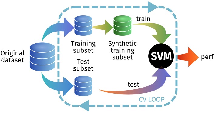
The rise of neuroimaging in research and clinical practice, together with the development of new machine learning techniques has strongly encouraged the Computer Aided Diagnosis (CAD) of different diseases and disorders. However, these algorithms are often tested in proprietary datasets to which the access is limited and, therefore, a direct comparison between CAD procedures is not possible. Furthermore, the sample size is often small for developing accurate machine learning methods. Multi-center initiatives are currently a very useful, although limited, tool in the recruitment of large populations and standardization of CAD evaluation. Conversely, we propose a brain image synthesis procedure intended to generate a new image set that share characteristics with an original one. Our system focuses on nuclear imaging modalities such as PET or SPECT brain images. We analyze the dataset by applying PCA to the original dataset, and then model the distribution of samples in the projected eigenbrain space using a Probability Density Function (PDF) estimator. Once the model has been built, we can generate new coordinates on the eigenbrain space belonging to the same class, which can be then projected back to the image space. The system has been evaluated on different functional neuroimaging datasets assessing the: resemblance of the synthetic images with the original ones, the differences between them, their generalization ability and the independence of the synthetic dataset with respect to the original. The synthetic images maintain the differences between groups found at the original dataset, with no significant differences when comparing them to real-world samples. Furthermore, they featured a similar performance and generalization capability to that of the original dataset. These results prove that these images are suitable for standardizing the evaluation of CAD pipelines, and providing data augmentation in machine learning systems -e.g. in deep learning-, or even to train future professionals at medical school.

摘要

神经影像学在研究和临床实践中的兴起,以及新机器学习技术的发展,极大地推动了不同疾病和病症的计算机辅助诊断(CAD)。然而,这些算法通常在访问受限的专有数据集中进行测试,因此无法直接比较CAD程序。此外,开发准确的机器学习方法时样本量往往较小。多中心倡议目前是招募大量人群和实现CAD评估标准化的非常有用但有限的工具。相反,我们提出了一种脑图像合成程序,旨在生成一组与原始图像具有共同特征的新图像。我们的系统专注于PET或SPECT脑图像等核成像模态。我们通过对原始数据集应用主成分分析(PCA)来分析数据集,然后使用概率密度函数(PDF)估计器对投影特征脑空间中的样本分布进行建模。一旦建立了模型,我们就可以在属于同一类别的特征脑空间上生成新的坐标,然后将其投影回图像空间。该系统已在不同的功能神经影像数据集上进行评估,评估内容包括:合成图像与原始图像的相似性、它们之间的差异、它们的泛化能力以及合成数据集相对于原始数据集的独立性。合成图像保留了原始数据集中发现的组间差异,与真实世界样本相比没有显著差异。此外,它们具有与原始数据集相似的性能和泛化能力。这些结果证明,这些图像适用于标准化CAD管道的评估,并在机器学习系统(例如深度学习)中提供数据增强,甚至用于培训医学院的未来专业人员。

https://cdn.ncbi.nlm.nih.gov/pmc/blobs/c7b4/5694626/6aa9823a2d25/fninf-11-00065-g0012.jpg
https://cdn.ncbi.nlm.nih.gov/pmc/blobs/c7b4/5694626/bad2538b0037/fninf-11-00065-g0001.jpg
https://cdn.ncbi.nlm.nih.gov/pmc/blobs/c7b4/5694626/a8d7cf1d9319/fninf-11-00065-g0002.jpg
https://cdn.ncbi.nlm.nih.gov/pmc/blobs/c7b4/5694626/d5fe3924ba2e/fninf-11-00065-g0003.jpg
https://cdn.ncbi.nlm.nih.gov/pmc/blobs/c7b4/5694626/833c710ac0c4/fninf-11-00065-g0004.jpg
https://cdn.ncbi.nlm.nih.gov/pmc/blobs/c7b4/5694626/33c04923f75a/fninf-11-00065-g0005.jpg
https://cdn.ncbi.nlm.nih.gov/pmc/blobs/c7b4/5694626/43e872b03d18/fninf-11-00065-g0006.jpg
https://cdn.ncbi.nlm.nih.gov/pmc/blobs/c7b4/5694626/ac2ae45d1e3e/fninf-11-00065-g0007.jpg
https://cdn.ncbi.nlm.nih.gov/pmc/blobs/c7b4/5694626/8ea4b4725a03/fninf-11-00065-g0008.jpg
https://cdn.ncbi.nlm.nih.gov/pmc/blobs/c7b4/5694626/5daac70c979a/fninf-11-00065-g0009.jpg
https://cdn.ncbi.nlm.nih.gov/pmc/blobs/c7b4/5694626/2b98dfca088d/fninf-11-00065-g0010.jpg
https://cdn.ncbi.nlm.nih.gov/pmc/blobs/c7b4/5694626/57fe7c9c9ea9/fninf-11-00065-g0011.jpg
https://cdn.ncbi.nlm.nih.gov/pmc/blobs/c7b4/5694626/6aa9823a2d25/fninf-11-00065-g0012.jpg

相似文献

1
Functional Brain Imaging Synthesis Based on Image Decomposition and Kernel Modeling: Application to Neurodegenerative Diseases.基于图像分解和核模型的功能性脑成像合成:在神经退行性疾病中的应用
Front Neuroinform. 2017 Nov 14;11:65. doi: 10.3389/fninf.2017.00065. eCollection 2017.
2
Three-Dimensional Eigenbrain for the Detection of Subjects and Brain Regions Related with Alzheimer's Disease.用于检测与阿尔茨海默病相关的受试者和脑区的三维特征脑
J Alzheimers Dis. 2016;50(4):1163-79. doi: 10.3233/JAD-150988.
3
Deep learning based pipelines for Alzheimer's disease diagnosis: A comparative study and a novel deep-ensemble method.基于深度学习的阿尔茨海默病诊断流程:一项对比研究及一种新型深度集成方法
Comput Biol Med. 2022 Feb;141:105032. doi: 10.1016/j.compbiomed.2021.105032. Epub 2021 Nov 21.
4
Detection of subjects and brain regions related to Alzheimer's disease using 3D MRI scans based on eigenbrain and machine learning.基于特征脑和机器学习,利用三维磁共振成像扫描检测与阿尔茨海默病相关的受试者和脑区。
Front Comput Neurosci. 2015 Jun 2;9:66. doi: 10.3389/fncom.2015.00066. eCollection 2015.
5
A Classification Algorithm by Combination of Feature Decomposition and Kernel Discriminant Analysis (KDA) for Automatic MR Brain Image Classification and AD Diagnosis.基于特征分解与核判别分析(KDA)组合的分类算法在自动磁共振脑图像分类与 AD 诊断中的应用。
Comput Math Methods Med. 2019 Dec 30;2019:1437123. doi: 10.1155/2019/1437123. eCollection 2019.
6
A parameter-efficient deep learning approach to predict conversion from mild cognitive impairment to Alzheimer's disease.一种参数高效的深度学习方法,用于预测轻度认知障碍向阿尔茨海默病的转化。
Neuroimage. 2019 Apr 1;189:276-287. doi: 10.1016/j.neuroimage.2019.01.031. Epub 2019 Jan 14.
7
Multi-Modality Cascaded Convolutional Neural Networks for Alzheimer's Disease Diagnosis.多模态级联卷积神经网络在阿尔茨海默病诊断中的应用。
Neuroinformatics. 2018 Oct;16(3-4):295-308. doi: 10.1007/s12021-018-9370-4.
8
BLENDS: Augmentation of Functional Magnetic Resonance Images for Machine Learning Using Anatomically Constrained Warping.BLENDS:基于解剖约束变形的机器学习功能磁共振图像增强。
Brain Connect. 2023 Mar;13(2):80-88. doi: 10.1089/brain.2021.0186. Epub 2022 Nov 4.
9
Learning using privileged information improves neuroimaging-based CAD of Alzheimer's disease: a comparative study.利用特权信息进行学习可提高基于神经影像学的阿尔茨海默病 CAD:一项比较研究。
Med Biol Eng Comput. 2019 Jul;57(7):1605-1616. doi: 10.1007/s11517-019-01974-3. Epub 2019 Apr 27.
10
Deep Learning and Machine Learning Algorithms for Retinal Image Analysis in Neurodegenerative Disease: Systematic Review of Datasets and Models.深度学习和机器学习算法在神经退行性疾病视网膜图像分析中的应用:数据集和模型的系统评价。
Transl Vis Sci Technol. 2024 Feb 1;13(2):16. doi: 10.1167/tvst.13.2.16.

引用本文的文献

1
The Major Hypotheses of Alzheimer's Disease: Related Nanotechnology-Based Approaches for Its Diagnosis and Treatment.阿尔茨海默病的主要假说:基于纳米技术的相关方法用于其诊断和治疗。
Cells. 2023 Nov 21;12(23):2669. doi: 10.3390/cells12232669.
2
Transfer Learning for Alzheimer's Disease through Neuroimaging Biomarkers: A Systematic Review.基于神经影像学生物标志物的阿尔茨海默病的迁移学习:系统综述。
Sensors (Basel). 2021 Oct 31;21(21):7259. doi: 10.3390/s21217259.
3
DiCyc: GAN-based deformation invariant cross-domain information fusion for medical image synthesis.

本文引用的文献

1
A review on neuroimaging-based classification studies and associated feature extraction methods for Alzheimer's disease and its prodromal stages.基于神经影像学的阿尔茨海默病及其前驱期分类研究及相关特征提取方法综述。
Neuroimage. 2017 Jul 15;155:530-548. doi: 10.1016/j.neuroimage.2017.03.057. Epub 2017 Apr 13.
2
Scanning the horizon: towards transparent and reproducible neuroimaging research.审视前沿:迈向透明且可重复的神经影像学研究。
Nat Rev Neurosci. 2017 Feb;18(2):115-126. doi: 10.1038/nrn.2016.167. Epub 2017 Jan 5.
3
Mini Mental State Examination and Logical Memory scores for entry into Alzheimer's disease trials.
DiCyc:基于生成对抗网络的变形不变跨域信息融合用于医学图像合成
Inf Fusion. 2021 Mar;67:147-160. doi: 10.1016/j.inffus.2020.10.015.
4
Deep learning in medical imaging and radiation therapy.深度学习在医学影像和放射治疗中的应用。
Med Phys. 2019 Jan;46(1):e1-e36. doi: 10.1002/mp.13264. Epub 2018 Nov 20.
用于纳入阿尔茨海默病试验的简易精神状态检查表和逻辑记忆评分。
Alzheimers Res Ther. 2016 Feb 22;8:9. doi: 10.1186/s13195-016-0176-z.
4
Building a FP-CIT SPECT Brain Template Using a Posterization Approach.使用色调分离方法构建FP-CIT单光子发射计算机断层扫描脑模板
Neuroinformatics. 2015 Oct;13(4):391-402. doi: 10.1007/s12021-015-9262-9.
5
Standardized evaluation of algorithms for computer-aided diagnosis of dementia based on structural MRI: the CADDementia challenge.基于结构磁共振成像的痴呆症计算机辅助诊断算法的标准化评估:CADDementia挑战赛
Neuroimage. 2015 May 1;111:562-79. doi: 10.1016/j.neuroimage.2015.01.048. Epub 2015 Jan 31.
6
Making big data open: data sharing in neuroimaging.使大数据开放:神经影像学中的数据共享。
Nat Neurosci. 2014 Nov;17(11):1510-7. doi: 10.1038/nn.3818. Epub 2014 Oct 28.
7
Big data from small data: data-sharing in the 'long tail' of neuroscience.从小数据到大数据:神经科学“长尾”中的数据共享。
Nat Neurosci. 2014 Nov;17(11):1442-7. doi: 10.1038/nn.3838.
8
Parametrization of textural patterns in 123I-ioflupane imaging for the automatic detection of Parkinsonism.用于帕金森病自动检测的123I-碘氟烷成像中纹理模式的参数化
Med Phys. 2014 Jan;41(1):012502. doi: 10.1118/1.4845115.
9
The autism brain imaging data exchange: towards a large-scale evaluation of the intrinsic brain architecture in autism.自闭症脑影像数据交换:走向自闭症内在大脑结构的大规模评估。
Mol Psychiatry. 2014 Jun;19(6):659-67. doi: 10.1038/mp.2013.78. Epub 2013 Jun 18.
10
Automatic assistance to Parkinson's disease diagnosis in DaTSCAN SPECT imaging.DaTSCAN SPECT 成像中帕金森病诊断的自动辅助。
Med Phys. 2012 Oct;39(10):5971-80. doi: 10.1118/1.4742055.