• 文献检索
  • 文档翻译
  • 深度研究
  • 学术资讯
  • Suppr Zotero 插件Zotero 插件
  • 邀请有礼
  • 套餐&价格
  • 历史记录
应用&插件
Suppr Zotero 插件Zotero 插件浏览器插件Mac 客户端Windows 客户端微信小程序
定价
高级版会员购买积分包购买API积分包
服务
文献检索文档翻译深度研究API 文档MCP 服务
关于我们
关于 Suppr公司介绍联系我们用户协议隐私条款
关注我们

Suppr 超能文献

核心技术专利:CN118964589B侵权必究
粤ICP备2023148730 号-1Suppr @ 2026

文献检索

告别复杂PubMed语法,用中文像聊天一样搜索,搜遍4000万医学文献。AI智能推荐,让科研检索更轻松。

立即免费搜索

文件翻译

保留排版,准确专业,支持PDF/Word/PPT等文件格式,支持 12+语言互译。

免费翻译文档

深度研究

AI帮你快速写综述,25分钟生成高质量综述,智能提取关键信息,辅助科研写作。

立即免费体验

新型小管间质性纤维化中周细胞-肌成纤维细胞转化的机制:核心岩藻糖基化调控。

Novel Mechanism of the Pericyte-Myofibroblast Transition in Renal Interstitial Fibrosis: Core Fucosylation Regulation.

机构信息

Department of Nephrology, The First Affiliated Hospital of Dalian Medical University, Key Laboratory of Kidney Disease of Liaoning Province, The Center for the Transformation Medicine of Kidney Disease of Liaoning Province, No. 222 Zhongshan Road, Dalian, 116011, China.

Department of Nephrology, Chinese PLA General Hospital, Chinese PLA Institute of Nephrology, State Key Laboratory of Kidney Diseases, National Clinical Research Center for Kidney Diseases, Fuxing Road 28, Haidian District, Beijing, 100853, China.

出版信息

Sci Rep. 2017 Dec 5;7(1):16914. doi: 10.1038/s41598-017-17193-5.

DOI:10.1038/s41598-017-17193-5
PMID:29209018
原文链接:https://pmc.ncbi.nlm.nih.gov/articles/PMC5717002/
Abstract

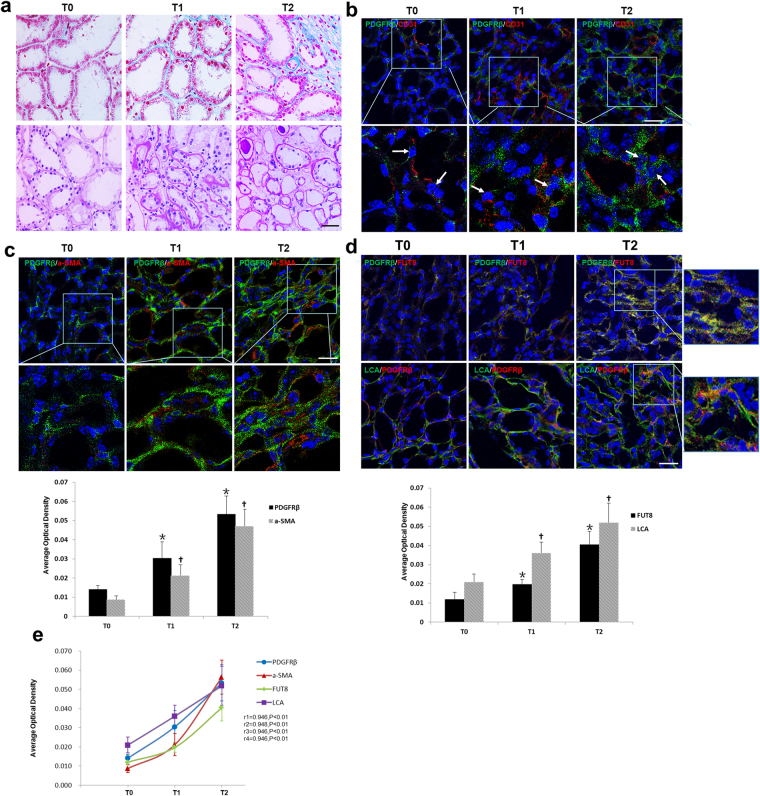
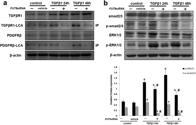
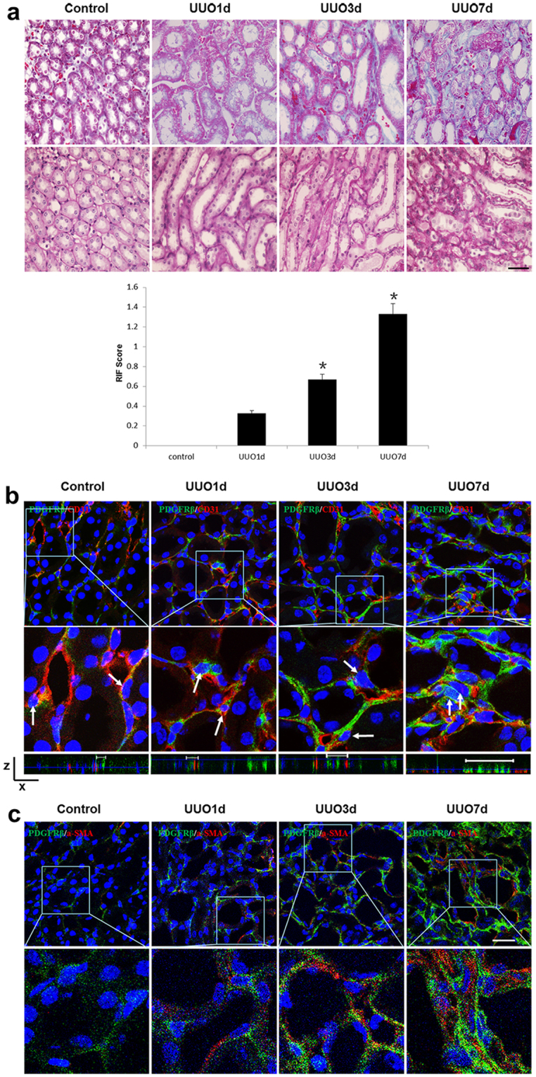
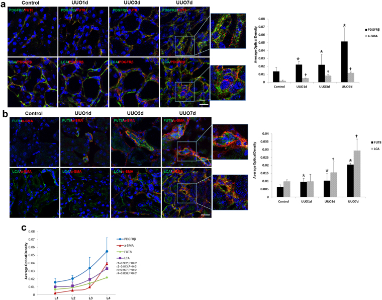
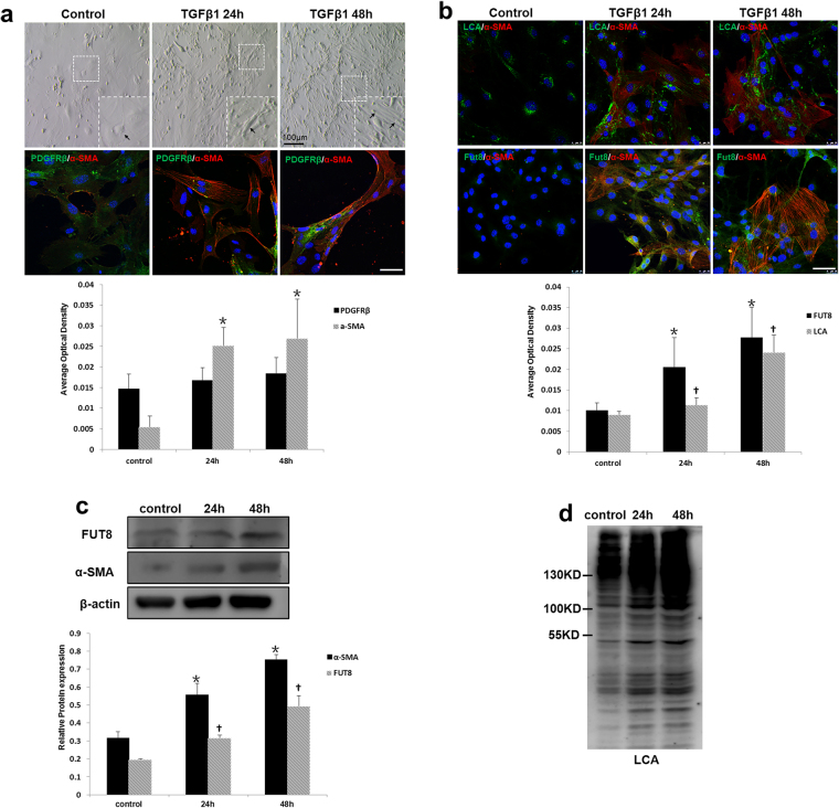
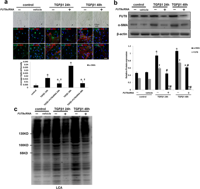
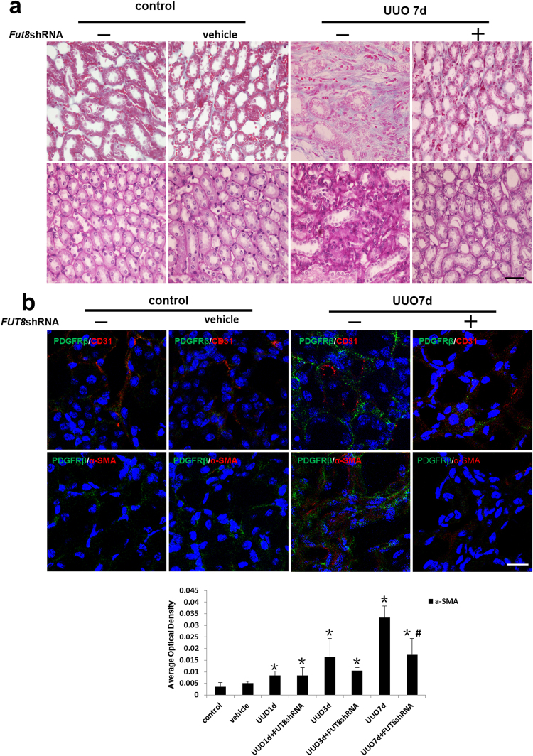
Pericytes have been identified as a major source of myofibroblasts in renal interstitial fibrosis (RIF). The overactivation of several signaling pathways, mainly the TGF-β and PDGF pathways, initiates the pericyte-myofibroblast transition during RIF. Key receptors in these two pathways have been shown to be modified by fucosyltransferase 8 (FUT8), the enzyme that catalyzes core fucosylation. This study postulated that core fucosylation might play an important role in regulating the pericyte transition in RIF. The data showed that core fucosylation increased with the extent of RIF in patients with IgA nephropathy (IgAN). Similarly, core fucosylation of pericytes increased in both a unilateral ureteral occlusion (UUO) mouse model and an in vitro model of pericyte transition. Inhibition of core fucosylation by adenoviral-mediated FUT8 shRNA in vivo and FUT8 siRNA in vitro significantly reduced pericyte transition and RIF. In addition, the activation of both the TGF-β/Smad and PDGF/ERK pathways was blocked by core fucosylation inhibition. In conclusion, core fucosylation may regulate the pericyte transition in RIF by modifying both the TGF-β/Smad and PDGF/ERK pathways. Glycosylation might be a novel "hub" target to prevent RIF.

摘要

周细胞已被确定为肾间质纤维化(RIF)中肌成纤维细胞的主要来源。在 RIF 期间,几种信号通路(主要是 TGF-β 和 PDGF 通路)的过度激活引发周细胞-肌成纤维细胞转化。这两条通路中的关键受体已被证明被岩藻糖基转移酶 8(FUT8)修饰,该酶催化核心岩藻糖基化。本研究假设核心岩藻糖基化可能在调节 RIF 中的周细胞转化中发挥重要作用。数据显示,在 IgA 肾病(IgAN)患者中,核心岩藻糖基化随着 RIF 的严重程度而增加。同样,在单侧输尿管梗阻(UUO)小鼠模型和周细胞转化的体外模型中,周细胞的核心岩藻糖基化均增加。体内通过腺病毒介导的 FUT8 shRNA 和体外 FUT8 siRNA 抑制核心岩藻糖基化显著减少了周细胞转化和 RIF。此外,核心岩藻糖基化抑制阻断了 TGF-β/Smad 和 PDGF/ERK 通路的激活。总之,核心岩藻糖基化可能通过修饰 TGF-β/Smad 和 PDGF/ERK 通路来调节 RIF 中的周细胞转化。糖基化可能是预防 RIF 的新型“枢纽”靶点。

https://cdn.ncbi.nlm.nih.gov/pmc/blobs/c6a7/5717002/7bbedb692cdb/41598_2017_17193_Fig7_HTML.jpg
https://cdn.ncbi.nlm.nih.gov/pmc/blobs/c6a7/5717002/351c20279d4f/41598_2017_17193_Fig1_HTML.jpg
https://cdn.ncbi.nlm.nih.gov/pmc/blobs/c6a7/5717002/33ddb59ffe4f/41598_2017_17193_Fig2_HTML.jpg
https://cdn.ncbi.nlm.nih.gov/pmc/blobs/c6a7/5717002/d7add7a4b06b/41598_2017_17193_Fig3_HTML.jpg
https://cdn.ncbi.nlm.nih.gov/pmc/blobs/c6a7/5717002/08cf7c03c40c/41598_2017_17193_Fig4_HTML.jpg
https://cdn.ncbi.nlm.nih.gov/pmc/blobs/c6a7/5717002/bd3ce6137818/41598_2017_17193_Fig5_HTML.jpg
https://cdn.ncbi.nlm.nih.gov/pmc/blobs/c6a7/5717002/7ddf06533e67/41598_2017_17193_Fig6_HTML.jpg
https://cdn.ncbi.nlm.nih.gov/pmc/blobs/c6a7/5717002/7bbedb692cdb/41598_2017_17193_Fig7_HTML.jpg

相似文献

1
Novel Mechanism of the Pericyte-Myofibroblast Transition in Renal Interstitial Fibrosis: Core Fucosylation Regulation.新型小管间质性纤维化中周细胞-肌成纤维细胞转化的机制:核心岩藻糖基化调控。
Sci Rep. 2017 Dec 5;7(1):16914. doi: 10.1038/s41598-017-17193-5.
2
Mechanisms of pulmonary fibrosis induced by core fucosylation in pericytes.周细胞中核心岩藻糖基化诱导肺纤维化的机制。
Int J Biochem Cell Biol. 2017 Jul;88:44-54. doi: 10.1016/j.biocel.2017.05.010. Epub 2017 May 5.
3
Inhibition of TGF-β1-receptor posttranslational core fucosylation attenuates rat renal interstitial fibrosis.抑制 TGF-β1 受体翻译后核心岩藻糖化可减轻大鼠肾间质纤维化。
Kidney Int. 2013 Jul;84(1):64-77. doi: 10.1038/ki.2013.82. Epub 2013 Mar 13.
4
Core fucosylation involvement in the paracrine regulation of proteinuria-induced renal interstitial fibrosis evaluated with the use of a microfluidic chip.利用微流控芯片评估核心岩藻糖基化在旁分泌调节蛋白尿诱导的肾间质纤维化中的作用。
Acta Biomater. 2022 Apr 1;142:99-112. doi: 10.1016/j.actbio.2022.02.020. Epub 2022 Feb 19.
5
Putative endothelial progenitor cells do not promote vascular repair but attenuate pericyte-myofibroblast transition in UUO-induced renal fibrosis.推定的内皮祖细胞不能促进血管修复,但可减轻 UUO 诱导的肾脏纤维化中的周细胞-肌成纤维细胞转化。
Stem Cell Res Ther. 2019 Mar 21;10(1):104. doi: 10.1186/s13287-019-1201-5.
6
The effect of core fucosylation-mediated regulation of multiple signaling pathways on lung pericyte activation and fibrosis.核心岩藻糖基化介导的多条信号通路调控对肺周细胞激活和纤维化的影响。
Int J Biochem Cell Biol. 2019 Dec;117:105639. doi: 10.1016/j.biocel.2019.105639. Epub 2019 Oct 24.
7
Inhibiting core fucosylation attenuates glucose-induced peritoneal fibrosis in rats.抑制核心岩藻糖基化可减轻葡萄糖诱导的大鼠腹膜纤维化。
Kidney Int. 2018 Jun;93(6):1384-1396. doi: 10.1016/j.kint.2017.12.023. Epub 2018 Mar 20.
8
Transforming growth factor β-1 stimulates profibrotic epithelial signaling to activate pericyte-myofibroblast transition in obstructive kidney fibrosis.转化生长因子β-1 刺激促纤维化上皮信号转导,激活阻塞性肾病纤维化中的周细胞-肌成纤维细胞转化。
Am J Pathol. 2013 Jan;182(1):118-31. doi: 10.1016/j.ajpath.2012.09.009. Epub 2012 Nov 9.
9
Novel insights into pericyte-myofibroblast transition and therapeutic targets in renal fibrosis.新颖的见解:周细胞-肌成纤维细胞转分化与肾纤维化治疗靶点。
J Formos Med Assoc. 2012 Nov;111(11):589-98. doi: 10.1016/j.jfma.2012.09.008. Epub 2012 Oct 24.
10
Platelet-derived growth factor receptor signaling activates pericyte-myofibroblast transition in obstructive and post-ischemic kidney fibrosis.血小板衍生生长因子受体信号激活阻塞性和缺血后肾脏纤维化中的周细胞-肌成纤维细胞转化。
Kidney Int. 2011 Dec;80(11):1170-81. doi: 10.1038/ki.2011.208. Epub 2011 Jun 29.

引用本文的文献

1
Glycosylation in kidney diseases.肾脏疾病中的糖基化作用。
Precis Clin Med. 2025 Jul 11;8(3):pbaf017. doi: 10.1093/pcmedi/pbaf017. eCollection 2025 Sep.
2
Effects of Oxygen Manipulation on Myofibroblast Phenotypic Transformation in Patients With Radiation-Induced Fibrosis.氧调控对放射性纤维化患者肌成纤维细胞表型转化的影响。
Wound Repair Regen. 2025 Jul-Aug;33(4):e70075. doi: 10.1111/wrr.70075.
3
Pericytes in the Pulmonary Microenvironment: Guardians or Adversaries?肺微环境中的周细胞:守护者还是对手?

本文引用的文献

1
Mechanisms of pulmonary fibrosis induced by core fucosylation in pericytes.周细胞中核心岩藻糖基化诱导肺纤维化的机制。
Int J Biochem Cell Biol. 2017 Jul;88:44-54. doi: 10.1016/j.biocel.2017.05.010. Epub 2017 May 5.
2
Urine Epidermal Growth Factor, Monocyte Chemoattractant Protein-1 or Their Ratio as Biomarkers for Interstitial Fibrosis and Tubular Atrophy in Primary Glomerulonephritis.尿表皮生长因子、单核细胞趋化蛋白-1或其比值作为原发性肾小球肾炎间质纤维化和肾小管萎缩的生物标志物
Kidney Blood Press Res. 2016;41(6):997-1007. doi: 10.1159/000452595. Epub 2016 Dec 19.
3
Progression of chronic kidney disease: too much cellular talk causes damage.
Lung. 2025 May 31;203(1):65. doi: 10.1007/s00408-025-00820-8.
4
Glycosylation in T2 high and Th17 Asthma: A Narrative Review.T2高型和Th17型哮喘中的糖基化:一篇叙述性综述
J Asthma Allergy. 2025 Apr 12;18:545-558. doi: 10.2147/JAA.S509940. eCollection 2025.
5
Role of microvascular pericyte dysfunction in antibody-mediated rejection following kidney transplantation.微血管周细胞功能障碍在肾移植后抗体介导性排斥反应中的作用
Ren Fail. 2025 Dec;47(1):2458749. doi: 10.1080/0886022X.2025.2458749. Epub 2025 Feb 5.
6
Cellular cross-talk drives mesenchymal transdifferentiation in diabetic kidney disease.细胞间相互作用驱动糖尿病肾病中的间充质转分化。
Front Med (Lausanne). 2025 Jan 7;11:1499473. doi: 10.3389/fmed.2024.1499473. eCollection 2024.
7
Fibroblast Heterogeneity in Inflammatory Bowel Disease.炎症性肠病中的成纤维细胞异质性
Int J Mol Sci. 2024 Dec 3;25(23):13008. doi: 10.3390/ijms252313008.
8
FUT8 upregulates CD36 and its core fucosylation to accelerate pericyte-myofibroblast transition through the mitochondrial-dependent apoptosis pathway during AKI-CKD.FUT8 通过上调 CD36 及其核心岩藻糖基化,通过线粒体依赖性凋亡通路在 AKI-CKD 期间加速周细胞-肌成纤维细胞转化。
Mol Med. 2024 Nov 20;30(1):222. doi: 10.1186/s10020-024-00994-6.
9
JR5558 mice are a reliable model to investigate subretinal fibrosis.JR5558 小鼠是研究视网膜下纤维化的可靠模型。
Sci Rep. 2024 Aug 13;14(1):18752. doi: 10.1038/s41598-024-66068-z.
10
Exploring unconventional targets in myofibroblast transdifferentiation outside classical TGF- signaling in renal fibrosis.探索肾纤维化中肌成纤维细胞转分化过程中经典转化生长因子信号通路之外的非常规靶点。
Front Physiol. 2024 May 14;15:1296504. doi: 10.3389/fphys.2024.1296504. eCollection 2024.
慢性肾脏病的进展:过多的细胞间交流导致损伤。
Kidney Int. 2017 Mar;91(3):552-560. doi: 10.1016/j.kint.2016.08.025. Epub 2016 Oct 20.
4
Human kidney pericytes produce renin.人肾周细胞产生肾素。
Kidney Int. 2016 Dec;90(6):1251-1261. doi: 10.1016/j.kint.2016.07.035. Epub 2016 Sep 24.
5
Risk factors for progression in children and young adults with IgA nephropathy: an analysis of 261 cases from the VALIGA European cohort.儿童和青年IgA肾病进展的危险因素:来自VALIGA欧洲队列的261例病例分析
Pediatr Nephrol. 2017 Jan;32(1):139-150. doi: 10.1007/s00467-016-3469-3. Epub 2016 Aug 25.
6
Silencing of microRNA-132 reduces renal fibrosis by selectively inhibiting myofibroblast proliferation.沉默 microRNA-132 可通过选择性抑制肌成纤维细胞增殖减少肾纤维化。
Kidney Int. 2016 Jun;89(6):1268-80. doi: 10.1016/j.kint.2016.01.029. Epub 2016 Apr 8.
7
TWEAK-Fn14 Signaling Activates Myofibroblasts to Drive Progression of Fibrotic Kidney Disease.肿瘤坏死因子样弱凋亡诱导因子-成纤维细胞生长因子诱导14信号通路激活肌成纤维细胞,推动纤维化肾病进展。
J Am Soc Nephrol. 2016 Dec;27(12):3639-3652. doi: 10.1681/ASN.2015111227. Epub 2016 Mar 29.
8
Gestation-associated changes in the glycosylation of placental insulin and insulin-like growth factor receptors.胎盘胰岛素及胰岛素样生长因子受体糖基化的妊娠相关变化。
Placenta. 2016 Mar;39:70-6. doi: 10.1016/j.placenta.2016.01.005. Epub 2016 Jan 8.
9
The role of PDGF-D in healthy and fibrotic kidneys.血小板衍生生长因子-D 在健康和纤维化肾脏中的作用。
Kidney Int. 2016 Apr;89(4):848-61. doi: 10.1016/j.kint.2015.12.037. Epub 2016 Feb 18.
10
Primary glomerulonephritides.原发性肾小球肾炎。
Lancet. 2016 May 14;387(10032):2036-48. doi: 10.1016/S0140-6736(16)00272-5. Epub 2016 Feb 25.