Departments of Nephrology, Tongji Hospital, Tongji Medical College, Huazhong University of Science and Technology, 1095 Jiefang Ave, Wuhan, 430030, Hubei, China.
Departments of Nutrition, Tongji Hospital, Tongji Medical College, Huazhong University of Science and Technology, 1095 Jiefang Ave, Wuhan, 430030, Hubei, China.
Stem Cell Res Ther. 2019 Mar 21;10(1):104. doi: 10.1186/s13287-019-1201-5.
Putative endothelial progenitor cells (pEPCs) have been confirmed to participate in alleviation of renal fibrosis in several ischaemic diseases. However, their mechanistic effect on renal fibrosis, which is characterized by vascular regression and further rarefaction-related pathology, remains unknown.
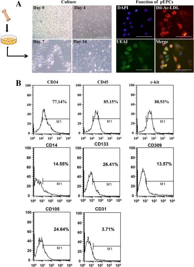
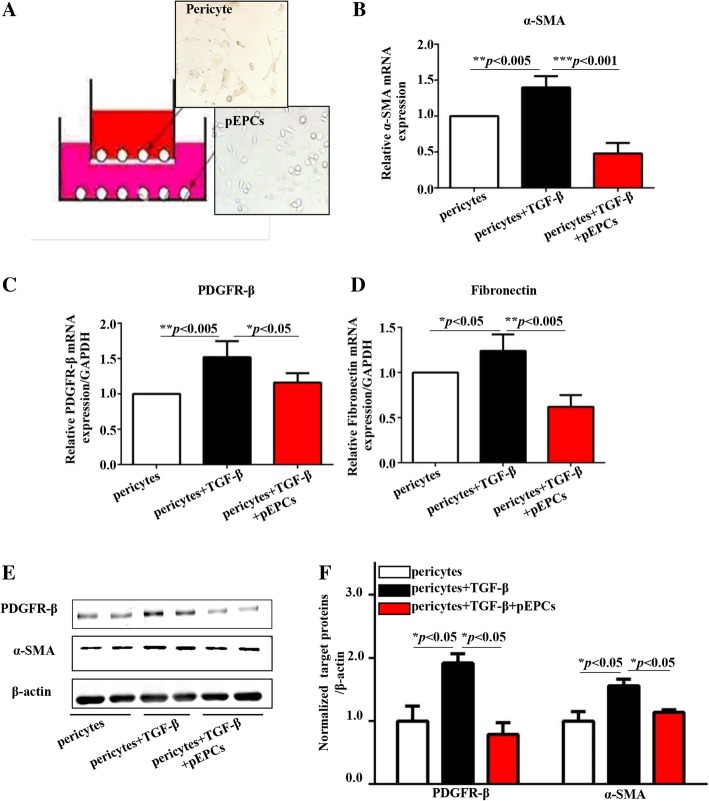
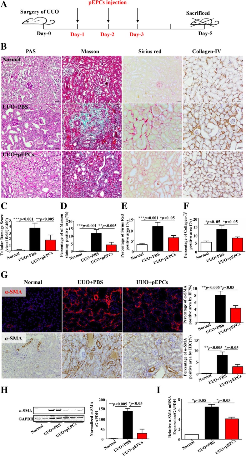
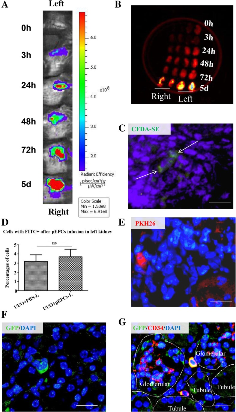
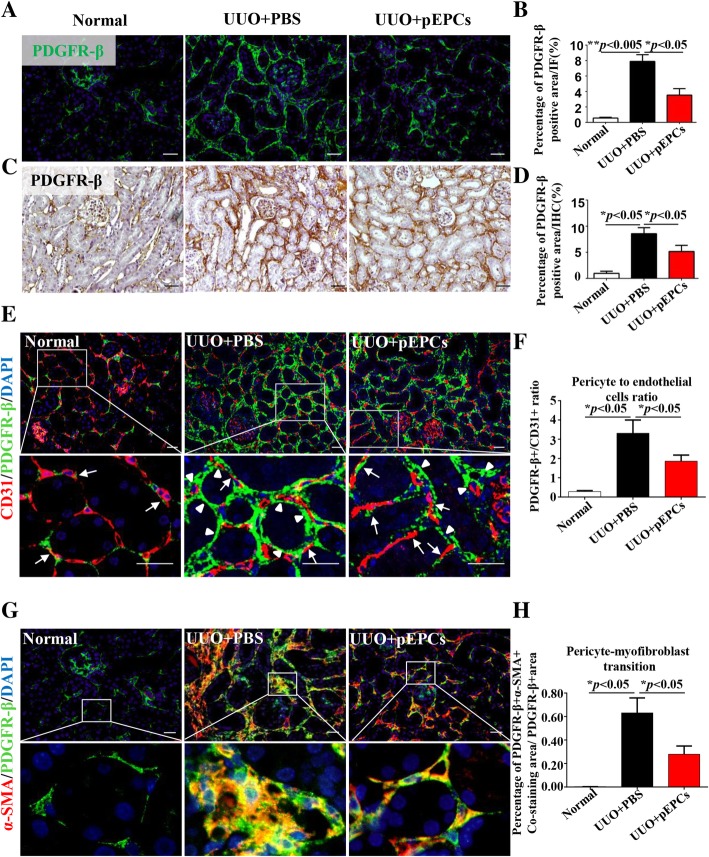
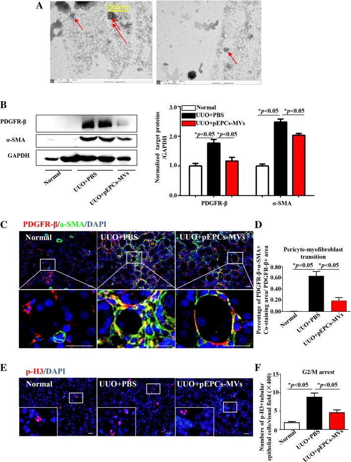
To explore the effect and molecular mechanisms by which pEPCs act on unilateral ureteral obstruction (UUO)-induced renal fibrosis, we isolated pEPCs from murine bone marrow. In vivo, pEPCs (2 × 10 cells/day) and pEPC-MVs (microvesicles) were injected into UUO mice via the tail vein. In vitro, pEPCs were co-cultured with renal-derived pericytes. Pericyte-myofibroblast transition was evaluated using the myofibroblast marker α-smooth muscle actin (α-SMA) and pericyte marker platelet-derived growth factor receptor β (PDGFR-β).
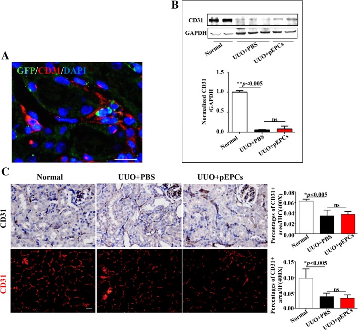
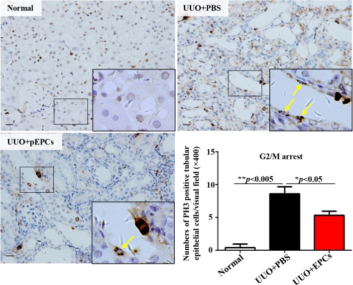
Exogenous supply of bone marrow-derived pEPCs attenuated renal fibrosis by decreasing pericyte-myofibroblast transition without significant vascular repair in the UUO model. Our results indicated that pEPCs regulated pericytes and their transition into myofibroblasts via pEPC-MVs. Co-culture of pericytes with pEPCs in vitro suggested that pEPCs inhibit transforming growth factor-β (TGF-β)-induced pericyte-myofibroblast transition via a paracrine pathway.
pEPCs effectively attenuated UUO-induced renal fibrosis by inhibiting pericyte-myofibroblast transition via a paracrine pathway, without promoting vascular repair.
已证实,假定的内皮祖细胞(pEPCs)可参与缓解几种缺血性疾病中的肾纤维化。然而,其对肾纤维化的机制作用(其特征为血管退化和进一步与稀疏相关的病理学)尚不清楚。
为了探讨 pEPCs 对单侧输尿管梗阻(UUO)诱导的肾纤维化的作用及其分子机制,我们从鼠骨髓中分离出 pEPCs。在体内,通过尾静脉将 pEPCs(2×10 个细胞/天)和 pEPC-MVs(微囊泡)注射到 UUO 小鼠体内。在体外,将 pEPCs 与肾源性周细胞共培养。通过肌成纤维细胞标志物 α-平滑肌肌动蛋白(α-SMA)和周细胞标志物血小板衍生生长因子受体 β(PDGFR-β)评估周细胞-肌成纤维细胞转化。
外源性骨髓来源的 pEPCs 通过减少周细胞-肌成纤维细胞转化,而在 UUO 模型中没有明显的血管修复,从而减轻了肾纤维化。我们的结果表明,pEPCs 通过 pEPC-MVs 调节周细胞及其向肌成纤维细胞的转化。体外周细胞与 pEPCs 共培养表明,pEPCs 通过旁分泌途径抑制转化生长因子-β(TGF-β)诱导的周细胞-肌成纤维细胞转化。
pEPCs 通过旁分泌途径有效抑制 UUO 诱导的肾纤维化,抑制周细胞-肌成纤维细胞转化,而不促进血管修复。