Biomedical Engineering Theme, Institute for Environment, Health and Societies, Brunel University London, Uxbridge, UB8 3PH, United Kingdom.
Department of Mechanical, Aerospace and Civil Engineering, College of Engineering, Design and Physical Sciences, Brunel University London, Uxbridge, UB8 3PH, United Kingdom.
J Biomed Mater Res A. 2018 Apr;106(4):1072-1081. doi: 10.1002/jbm.a.36308. Epub 2017 Dec 23.
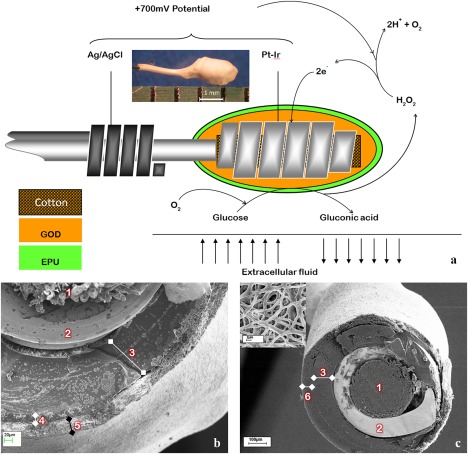
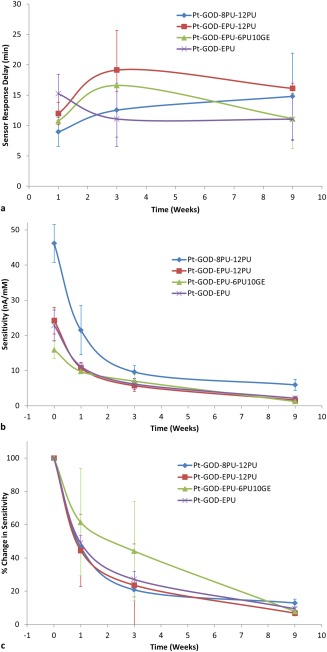
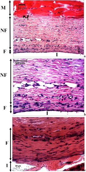
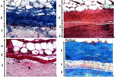
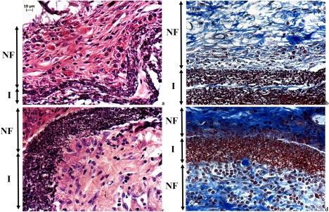
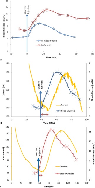
In vivo tissue responses and functional efficacy of electrospun membranes based on polyurethane (PU) and gelatin (GE) as biomimetic coatings for implantable glucose biosensors was investigated in a rat subcutaneous implantation model. Three electrospun membranes with optimized fiber diameters, pore sizes, and permeability, both single PU and coaxial PU-GE fibers and a solvent cast PU film were implanted in rats to evaluate tissue responses. For functional efficacy testing, four sensor variants coated with the above mentioned electrospun membranes as mass-transport limiting and outermost biomimetic coatings were implanted in rats. The electrospun PU membranes had micron sized pores that were not permeable to host cells when implanted in the body. However, PU-GE coaxial fiber membranes, having similar sized pores, were infiltrated with fibroblasts that deposited collagen in the membrane's pores. Such tissue response prevented the formation of dense fibrous capsule around the sensor coated with the PU-GE coaxial fiber membranes, which helped improve the in vivo sensitivity for at least 3 weeks compared to the traditional sensors in rat subcutaneous tissue. Furthermore, the better in vitro sensor's sensitivity due to electrospun PU as the mass-transport limiting membrane translated to better in vivo sensitivity. Thus, this study showed that electrospun membranes can play an important role in realizing long in vivo sensing lifetime of implantable glucose biosensors. © 2017 The Authors Journal of Biomedical Materials Research Part A Published by Wiley Periodicals, Inc. J Biomed Mater Res Part A: 106A: 1072-1081, 2018.
在大鼠皮下植入模型中,研究了基于聚氨酯(PU)和明胶(GE)的电纺膜作为植入式葡萄糖生物传感器仿生涂层的体内组织反应和功能效果。优化了纤维直径、孔径和渗透性的三种电纺膜,包括单 PU 和同轴 PU-GE 纤维以及溶剂浇铸的 PU 薄膜,被植入大鼠体内以评估组织反应。为了进行功能效果测试,将四种涂有上述电纺膜的传感器变体(作为质量传输限制和最外层仿生涂层)植入大鼠体内。当植入体内时,电纺 PU 膜具有微米大小的孔,这些孔对宿主细胞不可渗透。然而,具有相似孔径的 PU-GE 同轴纤维膜被成纤维细胞渗透,这些成纤维细胞在膜的孔中沉积胶原蛋白。这种组织反应阻止了传感器涂层周围形成致密的纤维囊,这有助于至少在 3 周内提高传感器的体内敏感性,与传统的大鼠皮下组织中的传感器相比。此外,由于电纺 PU 作为质量传输限制膜,体外传感器的敏感性更好,这转化为体内敏感性的提高。因此,本研究表明,电纺膜在实现植入式葡萄糖生物传感器的长期体内传感寿命方面可以发挥重要作用。©2017 作者 Wiley 期刊公司 生物材料研究杂志 A 部分:生物材料研究杂志 A 部分:106A:1072-1081,2018。