• 文献检索
  • 文档翻译
  • 深度研究
  • 学术资讯
  • Suppr Zotero 插件Zotero 插件
  • 邀请有礼
  • 套餐&价格
  • 历史记录
应用&插件
Suppr Zotero 插件Zotero 插件浏览器插件Mac 客户端Windows 客户端微信小程序
定价
高级版会员购买积分包购买API积分包
服务
文献检索文档翻译深度研究API 文档MCP 服务
关于我们
关于 Suppr公司介绍联系我们用户协议隐私条款
关注我们

Suppr 超能文献

核心技术专利:CN118964589B侵权必究
粤ICP备2023148730 号-1Suppr @ 2026

文献检索

告别复杂PubMed语法,用中文像聊天一样搜索,搜遍4000万医学文献。AI智能推荐,让科研检索更轻松。

立即免费搜索

文件翻译

保留排版,准确专业,支持PDF/Word/PPT等文件格式,支持 12+语言互译。

免费翻译文档

深度研究

AI帮你快速写综述,25分钟生成高质量综述,智能提取关键信息,辅助科研写作。

立即免费体验

在正常志愿者中使用光学相干断层扫描血管造影术对黄斑无血管区进行定量测量。

The quantitative measurements of foveal avascular zone using optical coherence tomography angiography in normal volunteers.

作者信息

Ghassemi Fariba, Mirshahi Reza, Bazvand Fatemeh, Fadakar Kaveh, Faghihi Houshang, Sabour Siamak

机构信息

Eye Research Center, Farabi Eye Hospital, Tehran University of Medical Sciences, Tehran, Iran.

Retina & Vitreous Service, Farabi Eye Hospital, Tehran University of Medical Sciences, Tehran, Iran.

出版信息

J Curr Ophthalmol. 2017 Jul 29;29(4):293-299. doi: 10.1016/j.joco.2017.06.004. eCollection 2017 Dec.

DOI:10.1016/j.joco.2017.06.004
PMID:29270477
原文链接:https://pmc.ncbi.nlm.nih.gov/articles/PMC5735238/
Abstract

PURPOSE

To provide normative data of foveal avascular zone (FAZ) and thickness.

METHODS

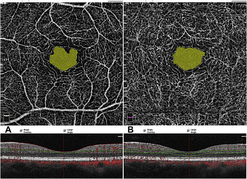
In this cross-sectional study both eyes of each normal subject were scanned with optical coherence tomography angiography (OCTA) for foveal superficial and deep avascular zone (FAZ) and central foveal thickness (CFT) and parafoveal thickness (PFT).

RESULTS

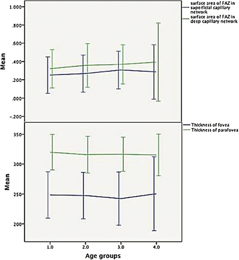
Out of a total of 224 eyes of 112 volunteers with a mean age of 37.03 (12-67) years, the mean superficial FAZ area was 0.27 mm, and deep FAZ area was 0.35 mm ( < 0.001), with no difference between both eyes. Females had a larger superficial (0.32 ± 0.11 mm versus 0.23 ± 0.09 mm) and deep FAZ (0.40 ± 0.14 mm versus 0.31 ± 0.10 mm) ( < 0.001) than males. By multivariate linear regression analysis, in normal eyes, superficial FAZ area varied significantly with the gender, CFT, and deep FAZ. Deep FAZ varied with the gender and CFT.

CONCLUSION

The gender and CFT influence the size of normal superficial and deep FAZ of capillary network.

摘要

目的

提供黄斑无血管区(FAZ)及其厚度的规范数据。

方法

在这项横断面研究中,使用光学相干断层扫描血管造影(OCTA)对每位正常受试者的双眼进行扫描,以测量黄斑浅层和深层无血管区(FAZ)、中心凹黄斑厚度(CFT)和旁中心凹厚度(PFT)。

结果

在112名平均年龄为37.03岁(12 - 67岁)的志愿者的224只眼中,浅层FAZ平均面积为0.27平方毫米,深层FAZ平均面积为0.35平方毫米(<0.001),双眼之间无差异。女性的浅层FAZ(0.32±0.11平方毫米对0.23±0.09平方毫米)和深层FAZ(0.40±0.14平方毫米对0.31±0.10平方毫米)均大于男性(<0.001)。通过多变量线性回归分析,在正常眼中,浅层FAZ面积随性别、CFT和深层FAZ有显著变化。深层FAZ随性别和CFT而变化。

结论

性别和CFT影响正常毛细血管网浅层和深层FAZ的大小。

https://cdn.ncbi.nlm.nih.gov/pmc/blobs/df6c/5735238/485ad1a29e99/gr2.jpg
https://cdn.ncbi.nlm.nih.gov/pmc/blobs/df6c/5735238/a6338a8978a6/gr1.jpg
https://cdn.ncbi.nlm.nih.gov/pmc/blobs/df6c/5735238/485ad1a29e99/gr2.jpg
https://cdn.ncbi.nlm.nih.gov/pmc/blobs/df6c/5735238/a6338a8978a6/gr1.jpg
https://cdn.ncbi.nlm.nih.gov/pmc/blobs/df6c/5735238/485ad1a29e99/gr2.jpg

相似文献

1
The quantitative measurements of foveal avascular zone using optical coherence tomography angiography in normal volunteers.在正常志愿者中使用光学相干断层扫描血管造影术对黄斑无血管区进行定量测量。
J Curr Ophthalmol. 2017 Jul 29;29(4):293-299. doi: 10.1016/j.joco.2017.06.004. eCollection 2017 Dec.
2
CORRELATION OF FOVEAL AVASCULAR ZONE SIZE WITH FOVEAL MORPHOLOGY IN NORMAL EYES USING OPTICAL COHERENCE TOMOGRAPHY ANGIOGRAPHY.使用光学相干断层扫描血管造影术对正常眼黄斑中心凹血管区大小与黄斑中心凹形态的相关性研究
Retina. 2015 Nov;35(11):2188-95. doi: 10.1097/IAE.0000000000000847.
3
Functional and Structural Outcomes at the Foveal Avascular Zone with Optical Coherence Tomography Following Macula off Retinal Detachment Repair.黄斑脱离视网膜脱离修复术后经光学相干断层扫描检测的黄斑无血管区的功能和结构转归
Clin Ophthalmol. 2020 Oct 13;14:3261-3270. doi: 10.2147/OPTH.S271944. eCollection 2020.
4
Analysis of foveal avascular zone by using spectral-domain optical coherence tomography angiography in healthy Indian eyes.利用频域光相干断层扫描血管造影分析健康印度人眼的中心凹无血管区。
Indian J Ophthalmol. 2024 Jun 1;72(6):838-843. doi: 10.4103/IJO.IJO_2212_23. Epub 2024 Mar 8.
5
VARIABILITY IN FOVEAL AVASCULAR ZONE AND CAPILLARY DENSITY USING OPTICAL COHERENCE TOMOGRAPHY ANGIOGRAPHY MACHINES IN HEALTHY EYES.使用光学相干断层扫描血管造影机检测健康眼睛的黄斑中心凹血管区和毛细血管密度的变异性
Retina. 2017 Nov;37(11):2102-2111. doi: 10.1097/IAE.0000000000001458.
6
[Correlation of capillary plexus with visual acuity in idiopathic macular epiretinal membrane eyes using optical coherence tomography angiography].[利用光学相干断层扫描血管造影术研究特发性黄斑视网膜前膜眼中毛细血管丛与视力的相关性]
Zhonghua Yan Ke Za Zhi. 2019 Oct 11;55(10):757-762. doi: 10.3760/cma.j.issn.0412-4081.2019.10.006.
7
Foveal Avascular Zone and Vessel Density in Healthy Subjects: An Optical Coherence Tomography Angiography Study.健康受试者的黄斑无血管区和血管密度:一项光学相干断层扫描血管造影研究。
J Ophthalmic Vis Res. 2018 Jul-Sep;13(3):260-265. doi: 10.4103/jovr.jovr_173_17.
8
Optical Coherence Tomography Angiography Evaluation of the Parafoveal Vasculature and Its Relationship With Ocular Factors.黄斑旁血管系统的光学相干断层扫描血管造影评估及其与眼部因素的关系。
Invest Ophthalmol Vis Sci. 2016 Jul 1;57(9):OCT224-34. doi: 10.1167/iovs.15-18869.
9
Interocular symmetry of the foveal avascular zone area in healthy eyes: a swept-source optical coherence tomography angiography study.健康眼中黄斑区无血管区面积的双眼对称性:扫频源光学相干断层血管造影研究。
Jpn J Ophthalmol. 2020 Mar;64(2):171-179. doi: 10.1007/s10384-020-00719-2. Epub 2020 Feb 3.
10
Foveal avascular zone in normal human eyes by optical coherence tomography angiography.光学相干断层扫描血管造影术观察正常人眼中心凹无血管区。
Photodiagnosis Photodyn Ther. 2023 Jun;42:103303. doi: 10.1016/j.pdpdt.2023.103303. Epub 2023 Jan 21.

引用本文的文献

1
The central retina vessel density and foveal avascular zone values of 792 healthy adults using optical coherence tomography angiography.792名健康成年人使用光学相干断层扫描血管造影术的中央视网膜血管密度和黄斑无血管区值。
Eye (Lond). 2024 Dec;38(18):3434-3442. doi: 10.1038/s41433-024-03320-w. Epub 2024 Sep 18.
2
Unlocking the Potential of Vessel Density and the Foveal Avascular Zone in Optical Coherence Tomography Angiography as Biomarkers in Alzheimer's Disease.揭示光学相干断层扫描血管造影中血管密度和黄斑无血管区作为阿尔茨海默病生物标志物的潜力。
Healthcare (Basel). 2024 Aug 9;12(16):1589. doi: 10.3390/healthcare12161589.
3

本文引用的文献

1
Optical Coherence Tomography Angiography Evaluation of the Parafoveal Vasculature and Its Relationship With Ocular Factors.黄斑旁血管系统的光学相干断层扫描血管造影评估及其与眼部因素的关系。
Invest Ophthalmol Vis Sci. 2016 Jul 1;57(9):OCT224-34. doi: 10.1167/iovs.15-18869.
2
Normative Data for Vascular Density in Superficial and Deep Capillary Plexuses of Healthy Adults Assessed by Optical Coherence Tomography Angiography.通过光学相干断层扫描血管造影评估健康成年人浅表和深部毛细血管丛血管密度的标准数据。
Invest Ophthalmol Vis Sci. 2016 Jul 1;57(9):OCT211-23. doi: 10.1167/iovs.15-18793.
3
Vascular Density in Retina and Choriocapillaris as Measured by Optical Coherence Tomography Angiography.
Using optical coherence tomography and optical coherence tomography angiography to delineate neurovascular homeostasis in migraine: a review.
利用光学相干断层扫描和光学相干断层扫描血管造影术描绘偏头痛中的神经血管稳态:综述
Front Neurosci. 2024 Apr 15;18:1376282. doi: 10.3389/fnins.2024.1376282. eCollection 2024.
4
Analysis of foveal avascular zone by using spectral-domain optical coherence tomography angiography in healthy Indian eyes.利用频域光相干断层扫描血管造影分析健康印度人眼的中心凹无血管区。
Indian J Ophthalmol. 2024 Jun 1;72(6):838-843. doi: 10.4103/IJO.IJO_2212_23. Epub 2024 Mar 8.
5
Quantitative Analysis of Inner, Middle, and Outer Retinal Thickness by Optical Coherence Tomography in Children and Adolescents.光学相干断层扫描对儿童和青少年视网膜内、中、外层厚度的定量分析
J Curr Ophthalmol. 2023 Dec 21;35(2):182-189. doi: 10.4103/joco.joco_141_23. eCollection 2023 Apr-Jun.
6
Evaluation of Choroidal and Retinal Features in Patients with Primary Vasculitis-An Original Optical Coherence Tomography and Optical Coherence Tomography Angiography Study.原发性血管炎患者脉络膜和视网膜特征评估——一项光学相干断层扫描和光学相干断层扫描血管造影的原创性研究
J Clin Med. 2023 Oct 29;12(21):6827. doi: 10.3390/jcm12216827.
7
Association of retinal microvascular abnormalities and neuromyelitis optica spectrum disorders with optical coherence tomography angiography.视网膜微血管异常和视神经脊髓炎谱系障碍与光学相干断层扫描血管造影的关联
Front Neurosci. 2023 Jun 9;17:1194661. doi: 10.3389/fnins.2023.1194661. eCollection 2023.
8
Retinal Optical Coherence Tomography Angiography Findings following Retinoblastoma Treatment by Chemotherapy.视网膜母细胞瘤化疗治疗后的视网膜光学相干断层扫描血管造影结果
J Curr Ophthalmol. 2023 Apr 29;34(4):452-459. doi: 10.4103/joco.joco_323_21. eCollection 2022 Oct-Dec.
9
Changes in Macular Thickness after Cataract Surgery in Patients with Open Angle Glaucoma.开角型青光眼患者白内障手术后黄斑厚度的变化
Diagnostics (Basel). 2023 Jan 9;13(2):244. doi: 10.3390/diagnostics13020244.
10
Quantification of Nonperfusion Area in Montaged Widefield OCT Angiography Using Deep Learning in Diabetic Retinopathy.在糖尿病视网膜病变中利用深度学习对拼接式超广角光学相干断层扫描血管造影中的无灌注区进行定量分析。
Ophthalmol Sci. 2021 May 12;1(2):100027. doi: 10.1016/j.xops.2021.100027. eCollection 2021 Jun.
光学相干断层扫描血管造影测量视网膜和脉络膜毛细血管的血管密度
Am J Ophthalmol. 2016 Aug;168:95-109. doi: 10.1016/j.ajo.2016.05.005. Epub 2016 May 14.
4
CORRELATION OF FOVEAL AVASCULAR ZONE SIZE WITH FOVEAL MORPHOLOGY IN NORMAL EYES USING OPTICAL COHERENCE TOMOGRAPHY ANGIOGRAPHY.使用光学相干断层扫描血管造影术对正常眼黄斑中心凹血管区大小与黄斑中心凹形态的相关性研究
Retina. 2015 Nov;35(11):2188-95. doi: 10.1097/IAE.0000000000000847.
5
ENLARGEMENT OF FOVEAL AVASCULAR ZONE IN DIABETIC EYES EVALUATED BY EN FACE OPTICAL COHERENCE TOMOGRAPHY ANGIOGRAPHY.通过正面光学相干断层扫描血管造影评估糖尿病患者眼部黄斑血管区扩大情况。
Retina. 2015 Nov;35(11):2377-83. doi: 10.1097/IAE.0000000000000849.
6
Measurement of Foveal Avascular Zone Dimensions and its Reliability in Healthy Eyes Using Optical Coherence Tomography Angiography.使用光学相干断层扫描血管造影术测量健康眼睛的黄斑无血管区尺寸及其可靠性
Am J Ophthalmol. 2016 Jan;161:50-5.e1. doi: 10.1016/j.ajo.2015.09.026. Epub 2015 Sep 28.
7
Reproducibility and repeatability of foveal avascular zone measurements in healthy subjects by optical coherence tomography angiography.光学相干断层扫描血管造影术测量健康受试者黄斑无血管区的可重复性和重复性
Br J Ophthalmol. 2016 May;100(5):671-6. doi: 10.1136/bjophthalmol-2015-307330. Epub 2015 Sep 16.
8
Optical coherence tomography angiography of the foveal avascular zone in diabetic retinopathy.糖尿病视网膜病变中黄斑无血管区的光学相干断层扫描血管造影
Graefes Arch Clin Exp Ophthalmol. 2016 Jun;254(6):1051-8. doi: 10.1007/s00417-015-3148-2. Epub 2015 Sep 4.
9
Quantitative Noninvasive Angiography of the Fovea Centralis Using Speckle Variance Optical Coherence Tomography.使用斑点方差光学相干断层扫描技术对中央凹进行定量无创血管造影。
Invest Ophthalmol Vis Sci. 2015 Aug;56(9):5074-86. doi: 10.1167/iovs.15-16773.
10
Noninvasive Visualization and Analysis of the Human Parafoveal Capillary Network Using Swept Source OCT Optical Microangiography.使用扫频源光学相干断层扫描光学微血管造影术对人黄斑旁毛细血管网络进行无创可视化和分析。
Invest Ophthalmol Vis Sci. 2015 Jun;56(6):3984-8. doi: 10.1167/iovs.15-16510.