Hao Tianpao, Chen Jingfeng, Zhi Shaoce, Zhang Qiyu, Chen Gang, Yu Fuxiang
Department of Hepatobiliary and Pancreatic Surgery, The First Affiliated Hospital, Wenzhou Medical University, Wenzhou, Zhejiang 325000, P.R. China.
Department of Anorectal Surgery, The Sixth Affiliated Hospital of Wenzhou Medical University, Lishui, Zhejiang 325000, P.R. China.
Exp Ther Med. 2017 Dec;14(6):5956-5964. doi: 10.3892/etm.2017.5333. Epub 2017 Oct 18.
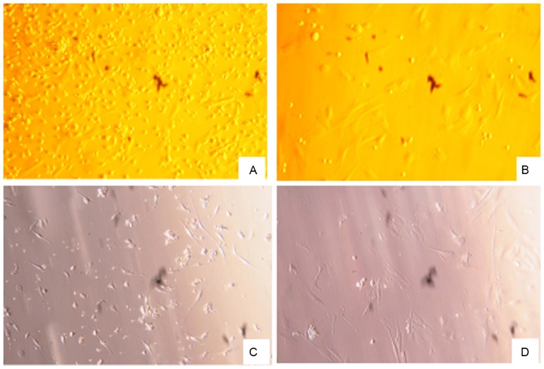
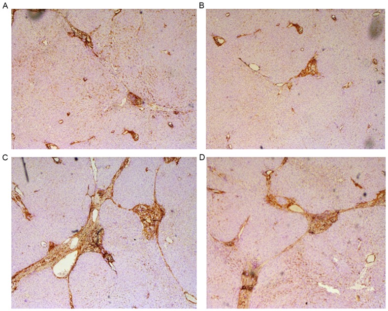
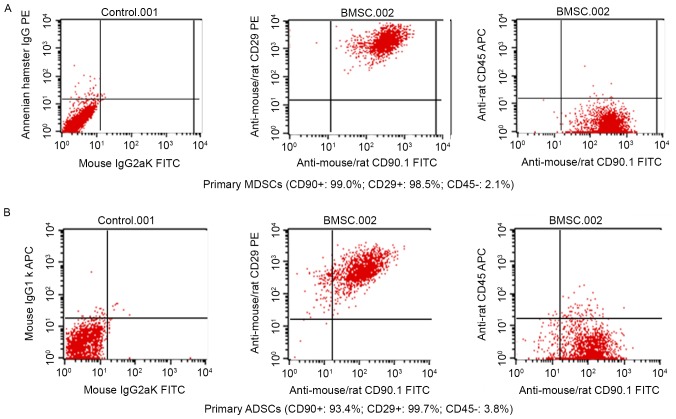
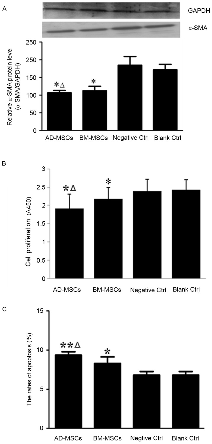
Mesenchymal stem cell (MSC) therapy has emerged as a potential novel method of treating liver fibrosis. To date, bone marrow-derived MSCs (BM-MSCs) and adipose tissue-derived MSCs (AD-MSCs) have not been analyzed with respect to their ability to combat liver fibrosis. The present study aimed to compare the capabilities of BM-MSCs and AD-MSCs in the treatment of liver fibrosis. BM-MSCs and AD-MSCs were taken from male Sprague-Dawley rats and cultured. Hepatic stellate cells (HSCs) were co-cultured with either BM-MSCs or AD-MSCs, and the effects of BM-MSCs or AD-MSCs on the proliferation, activation and apoptosis of HSCs were determined. The secretion of a selected group of cytokines by BM-MSCs and AD-MSCs was measured using enzyme-linked immunosorbent assays. Using a CCl-induced liver fibrosis animal model, the anti-inflammatory and anti-fibrotic effects of BM-MSCs or AD-MSCs against liver fibrosis were evaluated. The morphological examination and analysis of specific surface markers confirmed the successful preparation of BM-MSCs and AD-MSCs. Furthermore, the proliferation, activation and apoptosis of HSCs were significantly inhibited by BM-MSCs and AD-MSCs, with statistically greater reductions achieved by AD-MSCs compared with BM-MSCs. Direct comparison of the secretion of selected cytokines by BM-MSCs and AD-MSCs revealed that significantly higher levels of nerve growth factor and transforming growth factor-β1 were secreted in the AD-MSC culture medium, whereas levels of vascular endothelial growth factor and interleukin-10 did not differ significantly between AD-MSCs and BM-MSCs. studies using a CCl-induced liver fibrosis model demonstrated that inflammatory activity and fibrosis staging scores were significantly lower in the MSC-treated groups compared with controls. Although AD-MSCs improved anti-inflammatory and anti-fibrotic effects compared with BM-MSCs, these differences were not significant. Thus, the current study demonstrated that BM-MSCs and AD-MSCs are similarly effective at attenuating liver fibrosis by inhibiting the activation and proliferation of HSCs, as well as promoting the apoptosis of HSCs.
间充质干细胞(MSC)疗法已成为一种潜在的治疗肝纤维化的新方法。迄今为止,尚未对骨髓来源的间充质干细胞(BM-MSC)和脂肪组织来源的间充质干细胞(AD-MSC)对抗肝纤维化的能力进行分析。本研究旨在比较BM-MSC和AD-MSC在治疗肝纤维化方面的能力。从雄性Sprague-Dawley大鼠获取BM-MSC和AD-MSC并进行培养。将肝星状细胞(HSC)与BM-MSC或AD-MSC共培养,并测定BM-MSC或AD-MSC对HSC增殖、活化和凋亡的影响。使用酶联免疫吸附测定法测量BM-MSC和AD-MSC分泌的一组选定细胞因子。使用CCl诱导的肝纤维化动物模型,评估BM-MSC或AD-MSC对肝纤维化的抗炎和抗纤维化作用。特定表面标志物的形态学检查和分析证实成功制备了BM-MSC和AD-MSC。此外,BM-MSC和AD-MSC均显著抑制了HSC的增殖、活化和凋亡,与BM-MSC相比,AD-MSC在统计学上实现了更大程度的降低。对BM-MSC和AD-MSC分泌的选定细胞因子进行直接比较发现,AD-MSC培养基中神经生长因子和转化生长因子-β1的分泌水平显著更高,而AD-MSC和BM-MSC之间血管内皮生长因子和白细胞介素-10的水平没有显著差异。使用CCl诱导的肝纤维化模型的研究表明,与对照组相比,MSC治疗组的炎症活性和纤维化分期评分显著更低。尽管与BM-MSC相比,AD-MSC改善了抗炎和抗纤维化作用,但这些差异并不显著。因此,当前研究表明,BM-MSC和AD-MSC通过抑制HSC的活化和增殖以及促进HSC的凋亡,在减轻肝纤维化方面同样有效。