Marra Amanda N, Ulrich Marisa, White Audra, Springer Meghan, Wingert Rebecca A
Department of Biological Sciences, University of Notre Dame.
Department of Biological Sciences, University of Notre Dame;
J Vis Exp. 2017 Nov 18(129):56261. doi: 10.3791/56261.
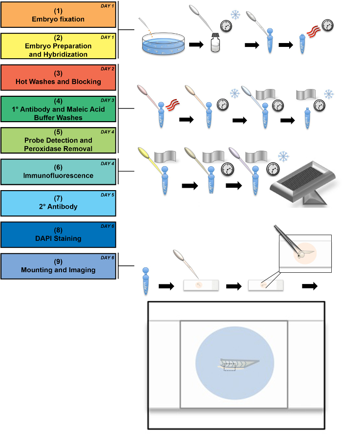
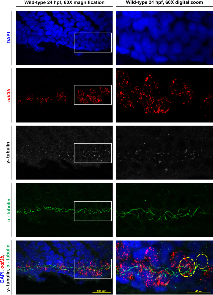
In recent years, the zebrafish embryo has emerged as a popular model to study developmental biology due to traits such as ex utero embryo development and optical transparency. In particular, the zebrafish embryo has become an important organism to study vertebrate kidney organogenesis as well as multiciliated cell (MCC) development. To visualize MCCs in the embryonic zebrafish kidney, we have developed a combined protocol of whole-mount fluorescent in situ hybridization (FISH) and whole mount immunofluorescence (IF) that enables high resolution imaging. This manuscript describes our technique for co-localizing RNA transcripts and protein as a tool to better understand the regulation of developmental programs through the expression of various lineage factors.
近年来,斑马鱼胚胎因其诸如胚胎在体外发育和光学透明等特性,已成为研究发育生物学的常用模型。特别是,斑马鱼胚胎已成为研究脊椎动物肾脏器官发生以及多纤毛细胞(MCC)发育的重要生物体。为了在斑马鱼胚胎肾脏中可视化多纤毛细胞,我们开发了一种全组织荧光原位杂交(FISH)和全组织免疫荧光(IF)相结合的方案,能够进行高分辨率成像。本手稿描述了我们将RNA转录本和蛋白质共定位的技术,作为一种工具,通过各种谱系因子的表达来更好地理解发育程序的调控。