Hill David R, Huang Sha, Tsai Yu-Hwai, Spence Jason R, Young Vincent B
Department of Internal Medicine, Division of Gastroenterology, University of Michigan;
Department of Internal Medicine, Division of Gastroenterology, University of Michigan.
J Vis Exp. 2017 Dec 18(130):56960. doi: 10.3791/56960.
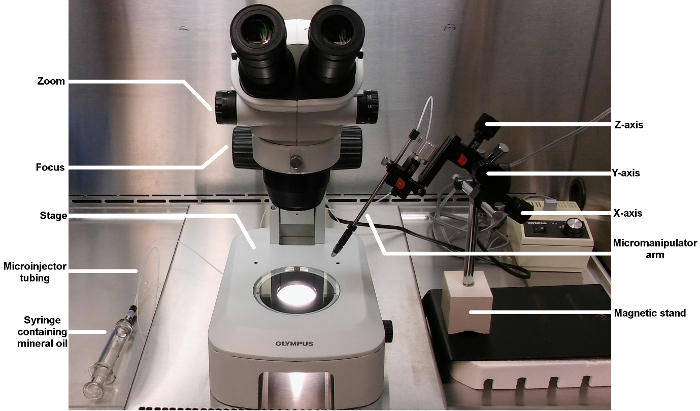
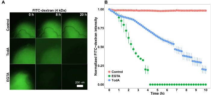
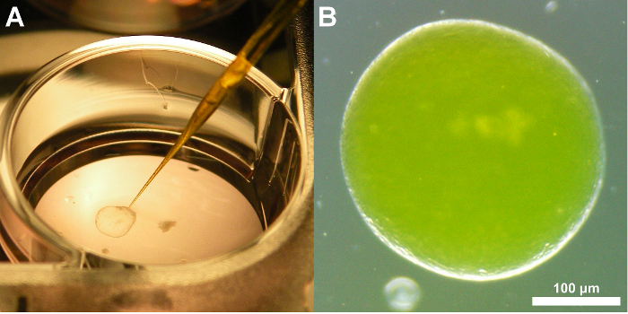
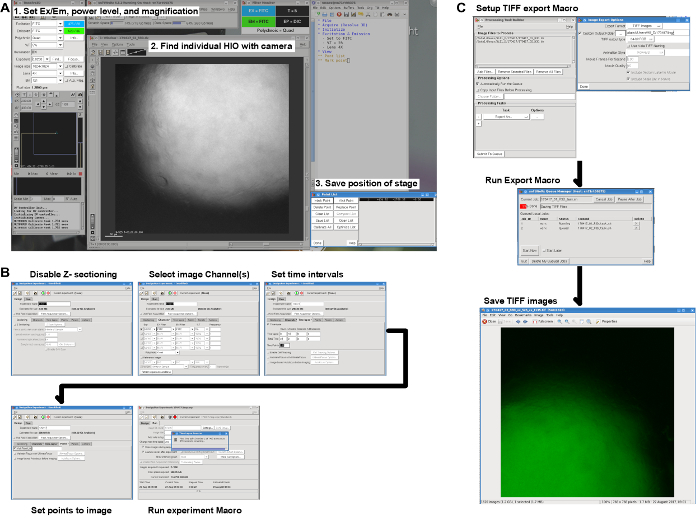
Advances in 3D culture of intestinal tissues obtained through biopsy or generated from pluripotent stem cells via directed differentiation, have resulted in sophisticated in vitro models of the intestinal mucosa. Leveraging these emerging model systems will require adaptation of tools and techniques developed for 2D culture systems and animals. Here, we describe a technique for measuring epithelial barrier permeability in human intestinal organoids in real-time. This is accomplished by microinjection of fluorescently-labeled dextran and imaging on an inverted microscope fitted with epifluorescent filters. Real-time measurement of the barrier permeability in intestinal organoids facilitates the generation of high-resolution temporal data in human intestinal epithelial tissue, although this technique can also be applied to fixed timepoint imaging approaches. This protocol is readily adaptable for the measurement of epithelial barrier permeability following exposure to pharmacologic agents, bacterial products or toxins, or live microorganisms. With minor modifications, this protocol can also serve as a general primer on microinjection of intestinal organoids and users may choose to supplement this protocol with additional or alternative downstream applications following microinjection.
通过活检获得或由多能干细胞经定向分化产生的肠道组织三维培养技术取得了进展,从而产生了复杂的肠黏膜体外模型。利用这些新兴的模型系统将需要对为二维培养系统和动物开发的工具和技术进行调整。在这里,我们描述了一种实时测量人肠道类器官上皮屏障通透性的技术。这是通过微量注射荧光标记的葡聚糖并在配备落射荧光滤光片的倒置显微镜上成像来实现的。实时测量肠道类器官中的屏障通透性有助于在人肠道上皮组织中生成高分辨率的时间数据,尽管该技术也可应用于固定时间点成像方法。该方案易于适用于测量暴露于药物、细菌产物或毒素或活微生物后的上皮屏障通透性。经过少量修改,该方案还可作为肠道类器官微量注射的通用入门指南,用户可以选择在微量注射后用其他或替代的下游应用来补充该方案。