Gupta Preeya K, Stevens Madelyn N, Kashyap Namita, Priestley Yos
Department of Ophthalmology, Duke University Eye Center, Durham, NC.
Duke University School of Medicine, Durham, NC.
Cornea. 2018 Apr;37(4):426-430. doi: 10.1097/ICO.0000000000001476.
To report the prevalence of meibomian gland atrophy and gland tortuosity in a pediatric population.
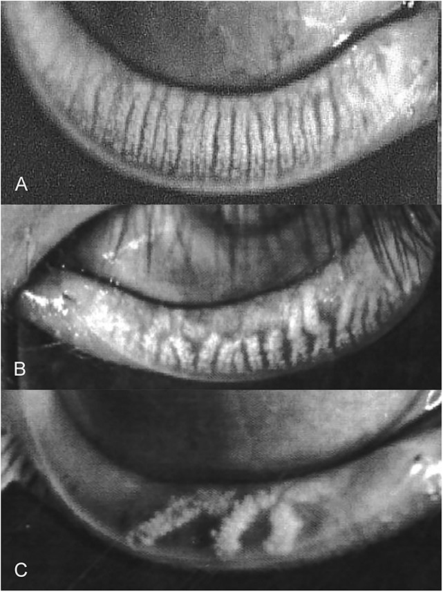
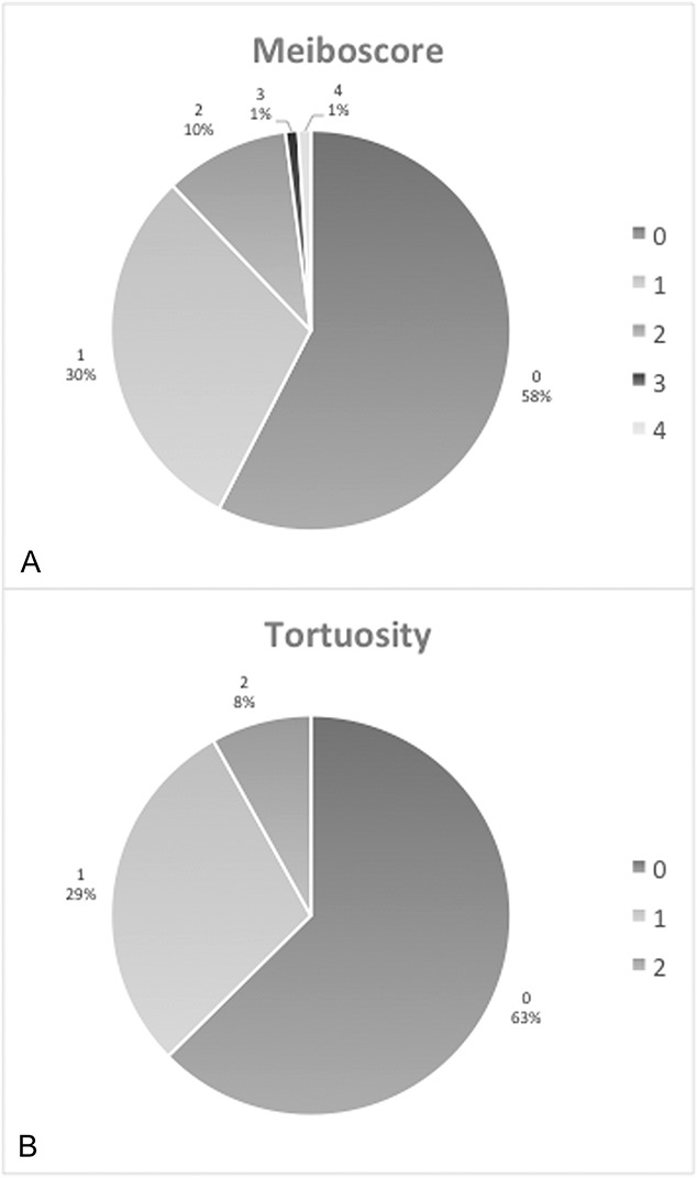
Participants who presented with no history of dry eye disease or meibomian gland dysfunction were recruited from the Duke University Eye Center. Meibography was performed and subjective symptoms were assessed through the Standard Patient Evaluation of Eye Dryness (SPEED) questionnaire. Grading of images was assessed by a masked rater using a previously validated 5-point meiboscale (0-4) for gland atrophy and a 3-point scale for gland tortuosity (0-2).
Ninety-nine eyes of 99 participants (50 females) aged 4 to 17 years (mean 9.6 years) were imaged. The mean meiboscore was 0.58 ± 0.80 (mean ± SD) for gland atrophy and 0.45 ± 0.64 for tortuosity. In all subjects, 42% (n = 42) had any evidence of meibomian gland atrophy (meiboscore >0) and 37% (n = 37) had any evidence of meibomian gland tortuosity. The majority of subjects had mild gland atrophy. No significant association was found between age, sex, or race and presence of gland atrophy. Males were significantly more likely to have gland tortuosity (P = 0.0124, odds ratio 3.36).
This study reveals a relatively high level of mild meibomian gland atrophy in the pediatric population, though moderate-severe gland atrophy was also present in this young population. This calls into question our current understanding of baseline gland architecture and suggests that perhaps clinicians should be examining young patients for meibomian gland atrophy and dysfunction because it may have implications for future development of dry eye disease.
报告儿科人群睑板腺萎缩和腺体扭曲的患病率。
从杜克大学眼科中心招募无干眼病史或睑板腺功能障碍病史的参与者。进行睑板腺造影,并通过干眼标准患者评估(SPEED)问卷评估主观症状。图像分级由一名盲态评分者使用先前验证的5分睑板腺评分量表(0 - 4)评估腺体萎缩情况,使用3分评分量表(0 - 2)评估腺体扭曲情况。
对99名年龄在4至17岁(平均9.6岁)的参与者(50名女性)的99只眼睛进行了成像。腺体萎缩的平均睑板腺评分为0.58±0.80(平均值±标准差),扭曲评分为0.45±0.64。在所有受试者中,42%(n = 42)有睑板腺萎缩的任何证据(睑板腺评分>0),37%(n = 37)有睑板腺扭曲的任何证据。大多数受试者有轻度腺体萎缩。在年龄、性别或种族与腺体萎缩的存在之间未发现显著关联。男性发生腺体扭曲的可能性显著更高(P = 0.0124,比值比3.36)。
本研究揭示儿科人群中轻度睑板腺萎缩的水平相对较高,尽管该年轻人群中也存在中度至重度腺体萎缩。这使我们对基线腺体结构的当前理解受到质疑,并表明或许临床医生应检查年轻患者是否存在睑板腺萎缩和功能障碍,因为这可能对干眼疾病的未来发展有影响。