Clinic for Diagnostic and Interventional Radiology, University Hospital Heidelberg, Heidelberg, Germany.
Clinic for Gynecology and Obstetrics, University Hospital Heidelberg, Heidelberg, Germany.
Theranostics. 2018 Jan 1;8(1):13-30. doi: 10.7150/thno.21089. eCollection 2018.
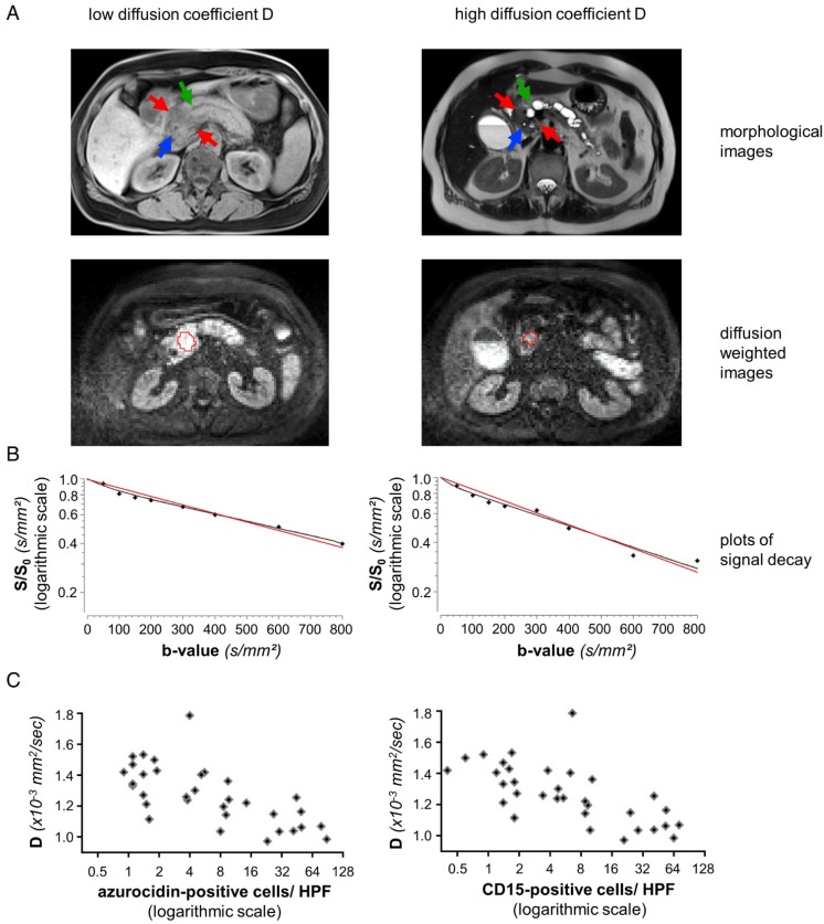
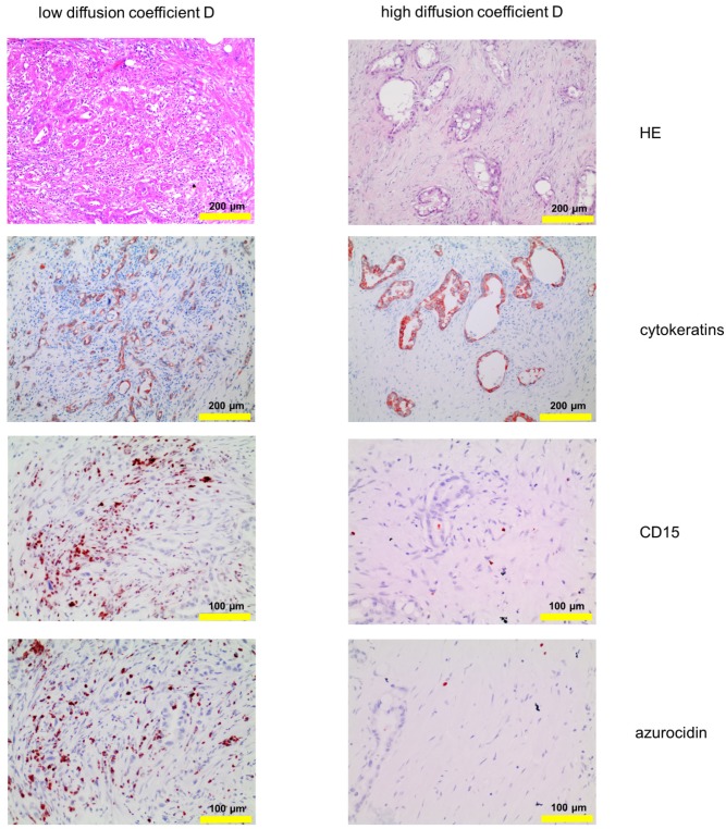
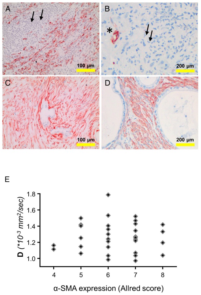
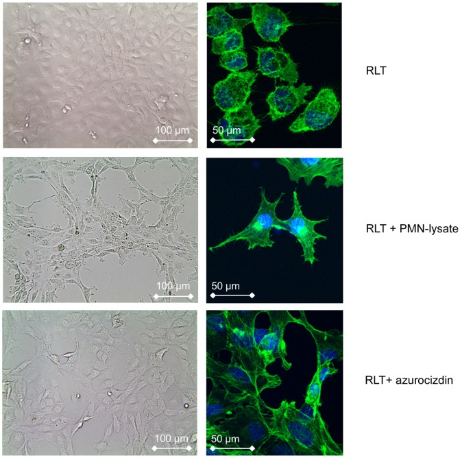
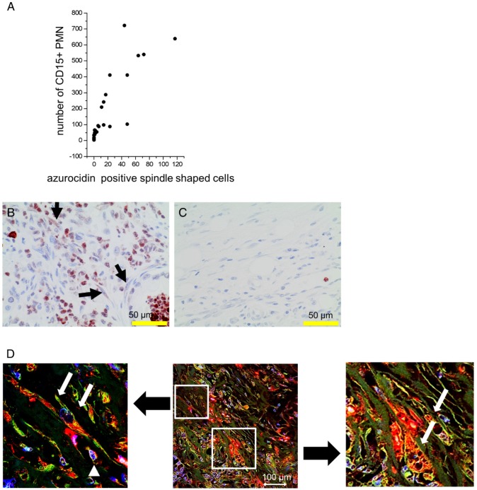
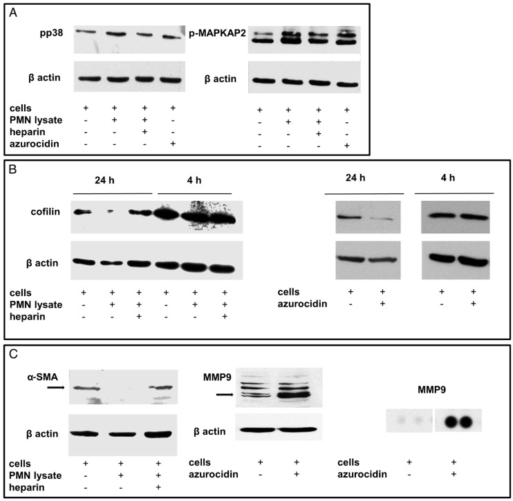
In pancreatic cancer (PDAC) intratumor infiltration of polymorphonuclear neutrophils (PMN) is associated with histologically apparent alterations of the tumor growth pattern. The aim of this study was to examine possible associations between PMN infiltration, tumor microarchitecture, and water diffusivity in diffusion-weighted magnetic resonance imaging (DW-MRI), and to further asses the underlying mechanisms. DW-MRI was performed in 33 PDAC patients prior to surgery. In parallel, tissue specimen were examined histologically for growth pattern, azurocidin-positive PMN infiltrates, and the presence of alpha-smooth muscle actin (α-SMA) and metalloproteinase 9 (MMP9)-positive myofibroblastic cells. For confirmation of the histological findings, a tissue microarray of a second cohort of patients (n=109) was prepared and examined similarly. For in vitro studies, the pancreatic stellate cell line RLT was co-cultivated either with isolated PMN, PMN-lysates, or recombinant azurocidin and characterized by Western blot, flow cytometry, and proteome profiler arrays. Tumors with high PMN density showed restricted water diffusion in DW-MRI and histologic apparent alterations of the tumor microarchitecture (microglandular, micropapillary, or overall poorly differentiated growth pattern) as opposed to tumors with scattered PMN. Areas with altered growth pattern lacked α-SMA-positive myofibroblastic cells. Tissue microarrays confirmed a close association of high PMN density with alterations of the tumor microarchitecture and revealed a significant association of high PMN density with poor histologic grade of differentiation (G3). In vitro experiments provided evidence for direct effects of PMN on stellate cells, where a change to a spindle shaped cell morphology in response to PMN and to PMN-derived azurocidin was seen. Azurocidin incorporated into stellate cells, where it associated with F-actin. Down-regulation of α-SMA was seen within hours, as was activation of the p38-cofilin axis, up-regulation of MMP9, and acquisition of intracellular lipid droplets, which together indicate a phenotype switch of the stellate cells. In PDAC, PMN infiltrates are associated with alterations of the tumor microarchitecture. As a causal relationship, we propose a reprogramming of stellate cells by PMN-derived azurocidin towards a phenotype, which affects the microarchitecture of the tumor.
在胰腺癌(PDAC)中,多形核中性粒细胞(PMN)的肿瘤内浸润与组织学上明显的肿瘤生长模式改变有关。本研究的目的是检查 PMN 浸润、肿瘤微结构和扩散加权磁共振成像(DW-MRI)中的水扩散之间可能存在的关联,并进一步评估潜在的机制。在手术前,对 33 名 PDAC 患者进行了 DW-MRI 检查。同时,对组织标本进行组织学检查,以确定生长模式、含azurocidin 的 PMN 浸润、α-平滑肌肌动蛋白(α-SMA)和金属蛋白酶 9(MMP9)阳性肌成纤维细胞的存在。为了确认组织学发现,准备了第二个患者队列(n=109)的组织微阵列并进行了类似的检查。在体外研究中,胰腺星状细胞系 RLT 与分离的 PMN、PMN 裂解物或重组 azurocidin 共培养,并通过 Western blot、流式细胞术和蛋白质组分析进行表征。PMN 密度高的肿瘤在 DW-MRI 中显示出受限的水扩散和组织学上明显的肿瘤微结构改变(微腺泡、微乳头状或整体分化不良的生长模式),而PMN 散在分布的肿瘤则相反。生长模式改变的区域缺乏α-SMA 阳性肌成纤维细胞。组织微阵列证实,PMN 密度与肿瘤微结构的改变密切相关,并显示PMN 密度与组织学分化程度差(G3)显著相关。体外实验提供了PMN 对星状细胞直接作用的证据,其中观察到星状细胞对 PMN 和 PMN 衍生的 azurocidin 的反应发生梭形细胞形态的改变。azurocidin 整合到星状细胞中,并与 F-肌动蛋白结合。在几小时内观察到α-SMA 的下调,同时还观察到 p38-丝切蛋白轴的激活、MMP9 的上调和细胞内脂滴的获得,这些共同表明星状细胞的表型转换。在 PDAC 中,PMN 浸润与肿瘤微结构的改变有关。作为一种因果关系,我们提出 PMN 衍生的 azurocidin 对星状细胞进行重编程,使其向影响肿瘤微结构的表型发展。