Garcia Erika Fernanda V, Loughin Catherine A, Marino Dominic J, Sackman Joseph, Umbaugh Scott E, Fu Jiyuan, Subedi Samrut, Lesser Martin L, Akerman Meredith, Schossler João Eduardo W
Department of Surgery, Long Island Veterinary Specialists, 163 South Service Road, Plainview, NY 11803, USA.
Computer Vision and Image Processing Laboratory, Electrical and Computer Engineering Department, Southern Illinois University at Edwardsville, Edwardsville, IL 62026-1801, USA.
Open Vet J. 2017;7(4):342-348. doi: 10.4314/ovj.v7i4.10. Epub 2017 Dec 8.
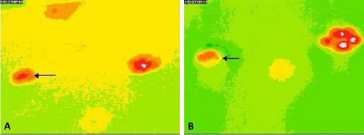
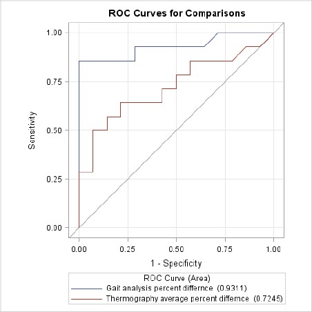
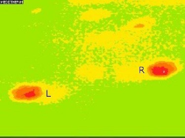
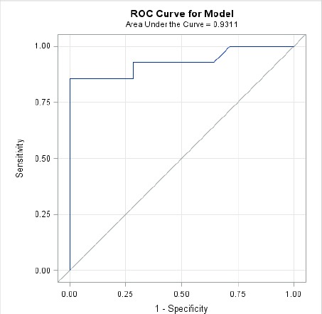
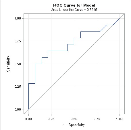
Subtle lameness makes it difficult to ascertain which is the affected limb. A study was conducted to investigate a change in the thermal pattern and temperature of the thermal image of the paw print in a lame pelvic limb compared to a non-lame pelvic limb of dogs confirmed by orthostatic analysis. Fourteen client owned dogs with a unilateral pelvic limb lameness and 14 healthy employee dogs were examined and the pelvic limbs radiographed. Thermal images of the paw print were taken after each dog was kept in a static position on a foam mat for 30 seconds. Average temperatures and thermographic patterns were analyzed. Analysis was performed in a static position. The asymmetry index for each stance variable and optimal cutoff point for the peak vertical force and thermal image temperatures were calculated. Image pattern analysis revealed 88% success in differentiating the lame group, and 100% in identifying the same thermal pattern in the healthy group. The mean of the peak vertical force revealed a 10.0% difference between the left and right pelvic limb in healthy dogs and a 72.4% between the lame and non-lame limb in the lame dog group. Asymmetry index analysis revealed 5% in the healthy group and 36.2% in the lame group. The optimal cutoff point for the peak vertical force to determine lameness was 41.77% (AUC = 0.93) and for MII 0.943% (AUC = 0.72). The results of this study highlight the change in the thermal pattern of the paw print in the lame pelvic limb compared to a non-lame pelvic limb in the lame group and the healthy group. Medical infrared imaging of the paw prints can be utilized to screen for the lame limb in dogs.
轻微跛行使得很难确定哪条腿是患肢。本研究旨在调查与通过体位分析确诊的非跛行犬的骨盆肢相比,跛行犬骨盆肢爪印热图像的热模式和温度变化。对14只单侧骨盆肢跛行的客户拥有的犬和14只健康的员工犬进行了检查,并对骨盆肢进行了X光检查。每只犬在泡沫垫上保持静止姿势30秒后,拍摄爪印的热图像。分析平均温度和热成像模式。在静止位置进行分析。计算每个姿势变量的不对称指数以及峰值垂直力和热图像温度的最佳截止点。图像模式分析显示,区分跛行组的成功率为88%,识别健康组相同热模式的成功率为100%。健康犬左右骨盆肢的峰值垂直力平均值相差10.0%,跛行犬组中跛行肢与非跛行肢之间相差72.4%。不对称指数分析显示,健康组为5%,跛行组为36.2%。确定跛行的峰值垂直力的最佳截止点为41.77%(AUC = 0.93),MII为0.943%(AUC = 0.72)。本研究结果突出了跛行组中跛行骨盆肢与非跛行骨盆肢相比爪印热模式的变化。爪印的医学红外成像可用于筛查犬的跛行肢。