Department of Pathology, China Medical University Hospital, Taichung, Taiwan.
Department of Radiation Oncology, China Medical University Hospital, Taichung, Taiwan.
Sci Rep. 2018 Jan 8;8(1):105. doi: 10.1038/s41598-017-18489-2.
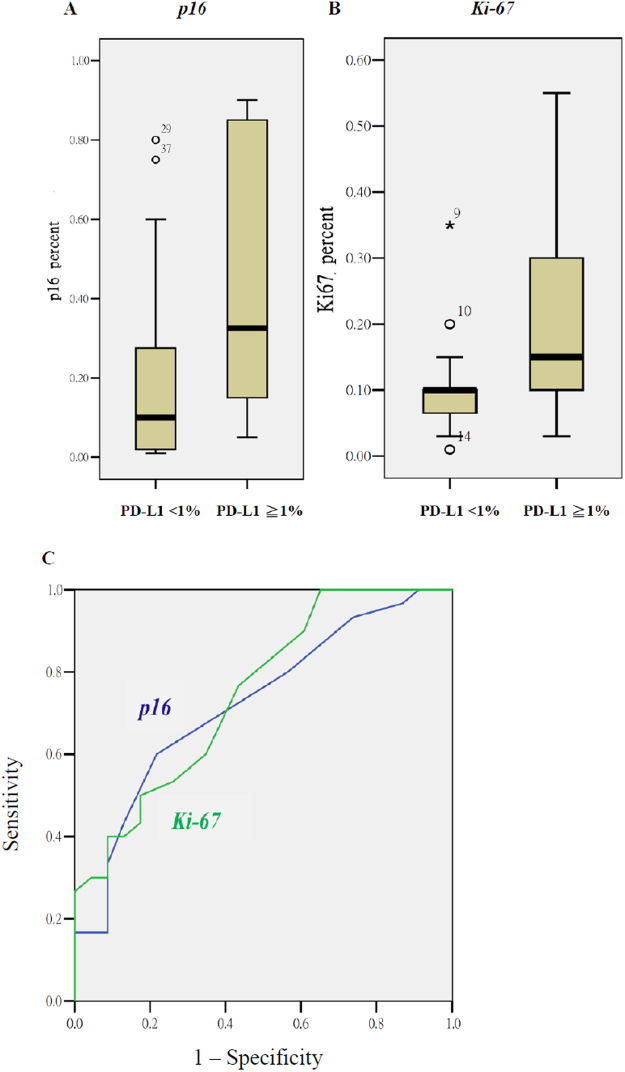
To know tumor PD-L1 expression through IHC or the FDG-PET related radiomics, we investigated the association between programmed cell death protein 1 ligand (PD-L1) expression and immunohistochemical (IHC) biomarkers or textural features of 18F-fluoro-2-deoxdeoxyglucose positron emission tomography (F-FDG PET) in 53 oropharyngeal or hypopharyngeal cancer patients who were ready to undergo radiotherapy-based treatment. Differences in textural features or biomarkers between tumors with and without PD-L1 expression were tested using a Mann-Whitney U test. The predicted values for PD-L1 expression were examined using logistic regression analysis. The mean percentages of tumor PD-L1 expression were 6.2 ± 13.5. Eighteen tumors had PD-L1 expression ≥5%, whereas 30 tumors ≥1%. Using a 5% cutoff, the p16 staining percentage and the textural index of correlation were two factors associated with PD-L1 expression. The odds ratios (ORs) were 17.00 (p = 0.028) and 0.009 (p = 0.015), respectively. When dichotomizing PD-L1 at 1%, the p16 and Ki-67 staining percentages were two predictors for PD-L1 expression with ORs of 11.41 (p = 0.035) and 757.77 (p = 0.045). p16 and Ki-67 staining percentages and several PET/CT-derived textural features can provide supplemental information to determine tumor PD-L1 expression in HNCs.
为了通过免疫组织化学(IHC)或 FDG-PET 相关的放射组学来了解肿瘤 PD-L1 的表达,我们研究了程序性死亡蛋白 1 配体(PD-L1)表达与 53 例准备接受基于放疗的治疗的口咽或下咽癌患者的免疫组织化学(IHC)生物标志物或 18F-氟-2-脱氧葡萄糖正电子发射断层扫描(F-FDG PET)纹理特征之间的关联。使用 Mann-Whitney U 检验测试 PD-L1 表达阳性和阴性肿瘤之间纹理特征或生物标志物的差异。使用逻辑回归分析检查 PD-L1 表达的预测值。肿瘤 PD-L1 表达的平均百分比为 6.2±13.5。18 个肿瘤 PD-L1 表达≥5%,30 个肿瘤 PD-L1 表达≥1%。使用 5%的截止值,p16 染色百分比和纹理相关指数是与 PD-L1 表达相关的两个因素。比值比(OR)分别为 17.00(p=0.028)和 0.009(p=0.015)。当将 PD-L1 分为 1%时,p16 和 Ki-67 染色百分比是 PD-L1 表达的两个预测因子,OR 分别为 11.41(p=0.035)和 757.77(p=0.045)。p16 和 Ki-67 染色百分比和几个 PET/CT 衍生的纹理特征可以提供补充信息,以确定 HNC 中肿瘤 PD-L1 的表达。