• 文献检索
  • 文档翻译
  • 深度研究
  • 学术资讯
  • Suppr Zotero 插件Zotero 插件
  • 邀请有礼
  • 套餐&价格
  • 历史记录
应用&插件
Suppr Zotero 插件Zotero 插件浏览器插件Mac 客户端Windows 客户端微信小程序
定价
高级版会员购买积分包购买API积分包
服务
文献检索文档翻译深度研究API 文档MCP 服务
关于我们
关于 Suppr公司介绍联系我们用户协议隐私条款
关注我们

Suppr 超能文献

核心技术专利:CN118964589B侵权必究
粤ICP备2023148730 号-1Suppr @ 2026

文献检索

告别复杂PubMed语法,用中文像聊天一样搜索,搜遍4000万医学文献。AI智能推荐,让科研检索更轻松。

立即免费搜索

文件翻译

保留排版,准确专业,支持PDF/Word/PPT等文件格式,支持 12+语言互译。

免费翻译文档

深度研究

AI帮你快速写综述,25分钟生成高质量综述,智能提取关键信息,辅助科研写作。

立即免费体验

压迫性视神经病变和正常眼压性青光眼的视盘旁血管密度:严重程度对照比较。

Peripapillary Vascular Density in Compressive Optic Neuropathy and Normal-Tension Glaucoma: A Severity-Controlled Comparison.

机构信息

Department of Ophthalmology, Samsung Medical Center, Sungkyunkwan University School of Medicine, Seoul, Korea.

Department of Neurosurgery, Samsung Medical Center, Sungkyunkwan University School of Medicine, Seoul, Korea.

出版信息

Invest Ophthalmol Vis Sci. 2023 Sep 1;64(12):10. doi: 10.1167/iovs.64.12.10.

DOI:10.1167/iovs.64.12.10
PMID:37672287
原文链接:https://pmc.ncbi.nlm.nih.gov/articles/PMC10484018/
Abstract

PURPOSE

To investigate the differences in peripapillary vessel density (VD) between compressive optic neuropathy (CON) and normal-tension glaucoma (NTG).

METHODS

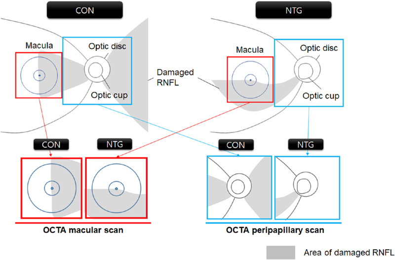
We compared patients with chronic CON and NTG, particularly after strictly controlling the mean extent of macular damage by the area of the ganglion cell-inner plexiform layer (GCIPL) loss in optical coherence tomography (OCT). We compared retinal nerve fiber layer (RNFL) and GCIPL thickness from OCT and peripapillary and macular VD from OCT angiography (OCTA) between the CON and NTG groups.

RESULTS

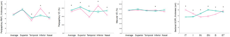
From the initial 184 patients with CON and 443 patients with OAG, we included 41 patients with CON (57 eyes) and 64 patients with NTG (75 eyes) with a comparable extent of macular GCIPL thinning. Under similar mean macular involvement, the peripapillary VD was significantly lower in the CON group than in the NTG group after considering the effects of age, spherical equivalent, visual field sensitivity, peripapillary RNFL (pRNFL) thickness, GCIPL thickness, and image quality scores (P < 0.001). Marked loss of VD in the temporal and nasal sectors in CON was notable, attributing to the significantly lower peripapillary VD compared to NTG.

CONCLUSIONS

Patients with CON had a significantly lower peripapillary VD than those with NTG under similar mean degrees of pRNFL thickness and GCIPL damage. Our results reveal the potential utility of OCTA VD besides OCT pRNFL thickness, in relation to different topographic patterns of pRNFL loss, and possible differences in the pathogenesis of microvascular compromise between CON and NTG.

摘要

目的

探讨压迫性视神经病变(CON)和正常眼压性青光眼(NTG)之间视乳头周围血管密度(VD)的差异。

方法

我们比较了慢性 CON 和 NTG 患者,特别是通过光学相干断层扫描(OCT)中神经节细胞-内丛状层(GCIPL)丢失面积来严格控制黄斑损伤的平均程度后。我们比较了 OCT 中的视网膜神经纤维层(RNFL)和 GCIPL 厚度以及 OCT 血管造影(OCTA)中的视乳头周围和黄斑 VD。

结果

从最初的 184 例 CON 患者和 443 例 OAG 患者中,我们纳入了 41 例 CON(57 只眼)和 64 例 NTG(75 只眼)患者,他们的黄斑 GCIPL 变薄程度相当。在考虑年龄、等效球镜、视野敏感度、视乳头周围 RNFL(pRNFL)厚度、GCIPL 厚度和图像质量评分的影响后,CON 组的视乳头周围 VD 明显低于 NTG 组(P<0.001)。在 CON 中,颞侧和鼻侧的 VD 明显丧失,这归因于与 NTG 相比,视乳头周围 VD 明显降低。

结论

在相似的平均 pRNFL 厚度和 GCIPL 损伤程度下,CON 患者的视乳头周围 VD 明显低于 NTG 患者。我们的研究结果揭示了 OCTA VD 除了 OCT pRNFL 厚度之外的潜在用途,与不同的 pRNFL 丧失的拓扑模式以及 CON 和 NTG 之间微血管损伤的发病机制可能存在差异有关。

https://cdn.ncbi.nlm.nih.gov/pmc/blobs/cbe0/10484018/049a308e96f9/iovs-64-12-10-f003.jpg
https://cdn.ncbi.nlm.nih.gov/pmc/blobs/cbe0/10484018/0fe803f3b1ba/iovs-64-12-10-f001.jpg
https://cdn.ncbi.nlm.nih.gov/pmc/blobs/cbe0/10484018/3fba9634291f/iovs-64-12-10-f002.jpg
https://cdn.ncbi.nlm.nih.gov/pmc/blobs/cbe0/10484018/049a308e96f9/iovs-64-12-10-f003.jpg
https://cdn.ncbi.nlm.nih.gov/pmc/blobs/cbe0/10484018/0fe803f3b1ba/iovs-64-12-10-f001.jpg
https://cdn.ncbi.nlm.nih.gov/pmc/blobs/cbe0/10484018/3fba9634291f/iovs-64-12-10-f002.jpg
https://cdn.ncbi.nlm.nih.gov/pmc/blobs/cbe0/10484018/049a308e96f9/iovs-64-12-10-f003.jpg

相似文献

1
Peripapillary Vascular Density in Compressive Optic Neuropathy and Normal-Tension Glaucoma: A Severity-Controlled Comparison.压迫性视神经病变和正常眼压性青光眼的视盘旁血管密度:严重程度对照比较。
Invest Ophthalmol Vis Sci. 2023 Sep 1;64(12):10. doi: 10.1167/iovs.64.12.10.
2
Correlation of Visual Field With Peripapillary Vessel Density Through Optical Coherence Tomography Angiography in Normal-Tension Glaucoma.正常眼压性青光眼中通过光学相干断层扫描血管造影术评估视野与视乳头周围血管密度的相关性
Transl Vis Sci Technol. 2020 Dec 17;9(13):26. doi: 10.1167/tvst.9.13.26. eCollection 2020 Dec.
3
Optical Coherence Tomography Angiography of Optic Disc in Eyes With Primary Open-angle Glaucoma and Normal-tension Glaucoma.原发性开角型青光眼和正常眼压性青光眼患者视盘的光学相干断层扫描血管造影。
J Glaucoma. 2019 Mar;28(3):243-251. doi: 10.1097/IJG.0000000000001184.
4
Discriminating Between Compressive Optic Neuropathy With Glaucoma-Like Cupping and Glaucomatous Optic Neuropathy Using OCT and OCTA.利用 OCT 和 OCTA 鉴别伴青光眼性杯盘比增大的压迫性视神经病变和青光眼性视神经病变。
Transl Vis Sci Technol. 2023 Mar 1;12(3):11. doi: 10.1167/tvst.12.3.11.
5
Effect of Nimodipine on Macular and Peripapillary Capillary Vessel Density in Patients with Normal-tension Glaucoma Using Optical Coherence Tomography Angiography.尼莫地平对正常眼压性青光眼患者用光学相干断层扫描血管造影测量的黄斑及视盘周围毛细血管密度的影响。
Curr Eye Res. 2021 Dec;46(12):1861-1866. doi: 10.1080/02713683.2021.1944645. Epub 2021 Jul 30.
6
Topographic comparison of the retinal microvascular changes between patients with compressive and glaucomatous optic neuropathies.压迫性和青光眼视神经病变患者视网膜微血管变化的地形比较。
Sci Rep. 2023 Dec 19;13(1):22569. doi: 10.1038/s41598-023-50068-6.
7
Comparison of the retinal microvasculature between compressive and glaucomatous optic neuropathy.比较压迫性和青光眼性视神经病变的视网膜微血管。
Graefes Arch Clin Exp Ophthalmol. 2023 Dec;261(12):3589-3597. doi: 10.1007/s00417-023-06137-7. Epub 2023 Jun 22.
8
Optical Coherence Tomography Angiography Vessel Density Changes in Normal-tension Glaucoma Treated With Carteolol, Brimonidine, or Dorzolamide.正常眼压性青光眼经卡替洛尔、溴莫尼定或多佐胺治疗后的光学相干断层扫描血管密度变化。
J Glaucoma. 2021 Aug 1;30(8):690-696. doi: 10.1097/IJG.0000000000001859.
9
Optical Coherence Tomography Angiography Macular and Peripapillary Vessel Perfusion Density in Healthy Subjects, Glaucoma Suspects, and Glaucoma Patients.光学相干断层扫描血管造影术检测健康受试者、青光眼疑似患者和青光眼患者黄斑及视乳头周围血管灌注密度
Invest Ophthalmol Vis Sci. 2017 Nov 1;58(13):5713-5722. doi: 10.1167/iovs.17-22865.
10
Optical Coherence Tomography Angiography Analysis of Peripapillary Choroidal Microvascular Density in Normal Tension Glaucoma and Primary Open Angle Glaucoma.正常眼压性青光眼和原发性开角型青光眼视乳头周围脉络膜微血管密度的光学相干断层扫描血管造影分析
J Glaucoma. 2025 Mar 1;34(3):189-197. doi: 10.1097/IJG.0000000000002499. Epub 2024 Sep 25.

引用本文的文献

1
Comparative of OCT and OCTA parameters in patients with early chronic angle-closure glaucoma and early pituitary adenoma.比较早期慢性闭角型青光眼和早期垂体腺瘤患者的 OCT 和 OCTA 参数。
Sci Rep. 2024 Sep 13;14(1):21448. doi: 10.1038/s41598-024-71103-0.

本文引用的文献

1
Discriminating Between Compressive Optic Neuropathy With Glaucoma-Like Cupping and Glaucomatous Optic Neuropathy Using OCT and OCTA.利用 OCT 和 OCTA 鉴别伴青光眼性杯盘比增大的压迫性视神经病变和青光眼性视神经病变。
Transl Vis Sci Technol. 2023 Mar 1;12(3):11. doi: 10.1167/tvst.12.3.11.
2
Change of Retinal Vessel Density After Lowering Intraocular Pressure in Ocular Hypertension.高眼压症患者降低眼压后视网膜血管密度的变化
Front Med (Lausanne). 2021 Dec 9;8:730327. doi: 10.3389/fmed.2021.730327. eCollection 2021.
3
Long-term reproducibility of optical coherence tomography angiography in healthy and stable glaucomatous eyes.
健康及稳定青光眼眼中光学相干断层扫描血管造影的长期可重复性。
Br J Ophthalmol. 2023 May;107(5):657-662. doi: 10.1136/bjophthalmol-2021-320034. Epub 2021 Dec 21.
4
Effect of algorithms and covariates in glaucoma diagnosis with optical coherence tomography angiography.光学相干断层扫描血管造影在青光眼诊断中算法和协变量的影响。
Br J Ophthalmol. 2022 Dec;106(12):1703-1709. doi: 10.1136/bjophthalmol-2020-318677. Epub 2021 Jun 28.
5
Changes in parafoveal and peripapillary perfusion after decompression surgery in chiasmal compression due to pituitary tumors.因垂体瘤导致的视交叉受压患者减压手术后旁中心区和视盘周围血流的变化。
Sci Rep. 2021 Feb 10;11(1):3464. doi: 10.1038/s41598-021-82151-1.
6
Correlation of Visual Field With Peripapillary Vessel Density Through Optical Coherence Tomography Angiography in Normal-Tension Glaucoma.正常眼压性青光眼中通过光学相干断层扫描血管造影术评估视野与视乳头周围血管密度的相关性
Transl Vis Sci Technol. 2020 Dec 17;9(13):26. doi: 10.1167/tvst.9.13.26. eCollection 2020 Dec.
7
Optical coherence tomography angiography in neuro-ophthalmology: Current clinical role and future perspectives.神经眼科中的光相干断层扫描血管造影:当前的临床作用和未来展望。
Surv Ophthalmol. 2021 May-Jun;66(3):471-481. doi: 10.1016/j.survophthal.2020.10.009. Epub 2020 Nov 3.
8
Longitudinal Changes in the Retinal Microstructures of Eyes With Chiasmal Compression.视交叉受压眼的视网膜微观结构的纵向变化。
Neurology. 2021 Jan 5;96(1):e131-e140. doi: 10.1212/WNL.0000000000011087. Epub 2020 Oct 22.
9
Glaucoma-like Parapapillary Choroidal Microvasculature Dropout in Patients with Compressive Optic Neuropathy.压迫性视神经病变患者的青光眼样视盘旁脉络膜小血管缺失。
Ophthalmology. 2020 Dec;127(12):1652-1662. doi: 10.1016/j.ophtha.2020.06.001. Epub 2020 Jun 7.
10
Artifacts and Anatomic Variations in Optical Coherence Tomography.光学相干断层扫描中的伪影和解剖变异。
Turk J Ophthalmol. 2020 Apr 29;50(2):99-106. doi: 10.4274/tjo.galenos.2019.78000.