Ogawa Madoka, Lester Robert, Akima Hiroshi, Gorgey Ashraf S
Graduate School of Education & Human Development, Nagoya University, Nagoya; Society for Promotion of Science, Tokyo, Japan.
Spinal Cord Injury and Disorders Center, Hunter Holmes McGuire VAMC, Richmond, VA, USA.
Neural Regen Res. 2017 Dec;12(12):2100-2105. doi: 10.4103/1673-5374.221170.
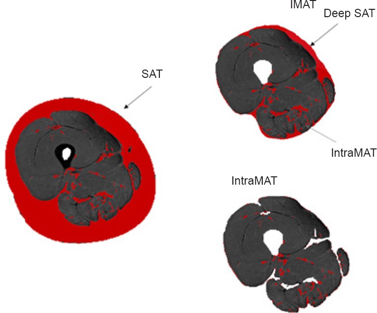
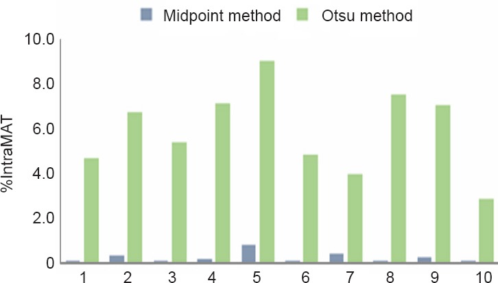
Ectopic adiposity has gained considerable attention because of its tight association with metabolic and cardiovascular health in persons with spinal cord injury (SCI). Ectopic adiposity is characterized by the storage of adipose tissue in non-subcutaneous sites. Magnetic resonance imaging (MRI) has proven to be an effective tool in quantifying ectopic adiposity and provides the opportunity to measure different adipose depots including intermuscular adipose tissue (IMAT) and intramuscular adipose tissue (IntraMAT) or intramuscular fat (IMF). It is highly important to distinguish and clearly define these compartments, because controversy still exists on how to accurately quantify these adipose depots. Investigators have relied on separating muscle from fat pixels based on their characteristic signal intensities. A common technique is plotting a threshold histogram that clearly separates between muscle and fat peaks. The cut-offs to separate between muscle and fat peaks are still not clearly defined and different cut-offs have been identified. This review will outline and compare the Midpoint and Otsu techniques, two methods used to determine the threshold between muscle and fat pixels on T1 weighted MRI. The process of water/fat segmentation using the Dixon method will also be outlined. We are hopeful that this review will trigger more research towards accurately quantifying ectopic adiposity due to its high relevance to cardiometabolic health after SCI.
异位脂肪沉积因其与脊髓损伤(SCI)患者的代谢和心血管健康密切相关而备受关注。异位脂肪沉积的特征是脂肪组织在非皮下部位储存。磁共振成像(MRI)已被证明是量化异位脂肪沉积的有效工具,并且提供了测量不同脂肪库的机会,包括肌间脂肪组织(IMAT)、肌内脂肪组织(IntraMAT)或肌内脂肪(IMF)。区分并明确定义这些区域非常重要,因为在如何准确量化这些脂肪库方面仍存在争议。研究人员依靠根据肌肉和脂肪像素的特征信号强度将它们分离。一种常用技术是绘制阈值直方图,以清晰区分肌肉和脂肪峰值。肌肉和脂肪峰值之间的分割阈值仍未明确界定,并且已确定了不同的分割阈值。本综述将概述并比较中点法和大津法这两种用于确定T1加权MRI上肌肉和脂肪像素之间阈值的方法。还将概述使用迪克森法进行水/脂肪分割的过程。我们希望本综述将引发更多关于准确量化异位脂肪沉积的研究,因为它与脊髓损伤后的心脏代谢健康高度相关。