Constantinides Christakis, Maguire Mahon, McNeill Eileen, Carnicer Ricardo, Swider Edyta, Srinivas Mangala, Carr Carolyn A, Schneider Jurgen E
Departments of Cardiovascular Medicine, Radcliffe Department of Medicine, University of Oxford, Oxford, United Kingdom.
Department of Tumor Immunology, Radboud University Medical Center, Radboud University, Nijmegen, The Netherlands.
PLoS One. 2018 Jan 11;13(1):e0190558. doi: 10.1371/journal.pone.0190558. eCollection 2018.
To a) achieve cardiac 19F-Magnetic Resonance Imaging (MRI) of perfluoro-crown-ether (PFCE) labeled cardiac progenitor stem cells (CPCs) and bone-derived bone marrow macrophages, b) determine label concentration and cellular load limits, and c) achieve spectroscopic and image-based quantification.
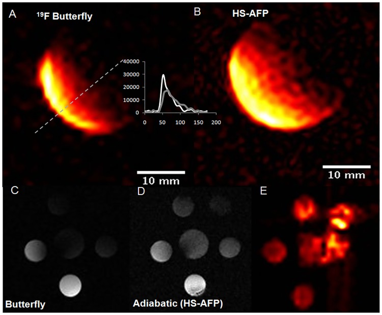
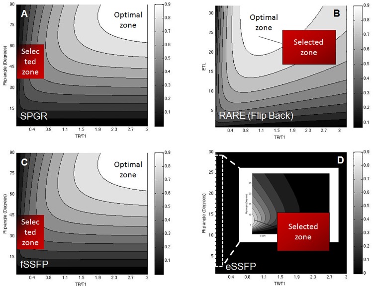
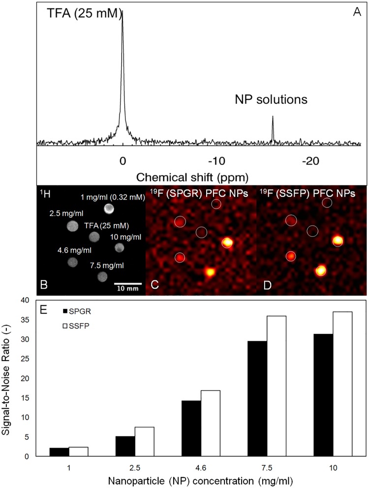
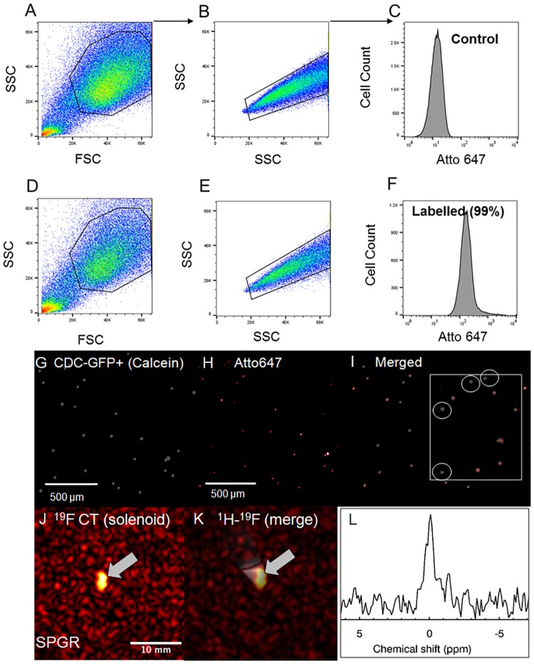
Theoretical simulations and experimental comparisons of spoiled-gradient echo (SPGR), rapid acquisition with relaxation enhancement (RARE), and steady state at free precession (SSFP) pulse sequences, and phantom validations, were conducted using 19F MRI/Magnetic Resonance Spectroscopy (MRS) at 9.4 T. Successful cell labeling was confirmed using flow cytometry and confocal microscopy. For CPC and macrophage concentration quantification, in vitro and post-mortem cardiac validations were pursued with the use of the transfection agent FuGENE. Feasibility of fast imaging is demonstrated in murine cardiac acquisitions in vivo, and in post-mortem murine skeletal and cardiac applications.
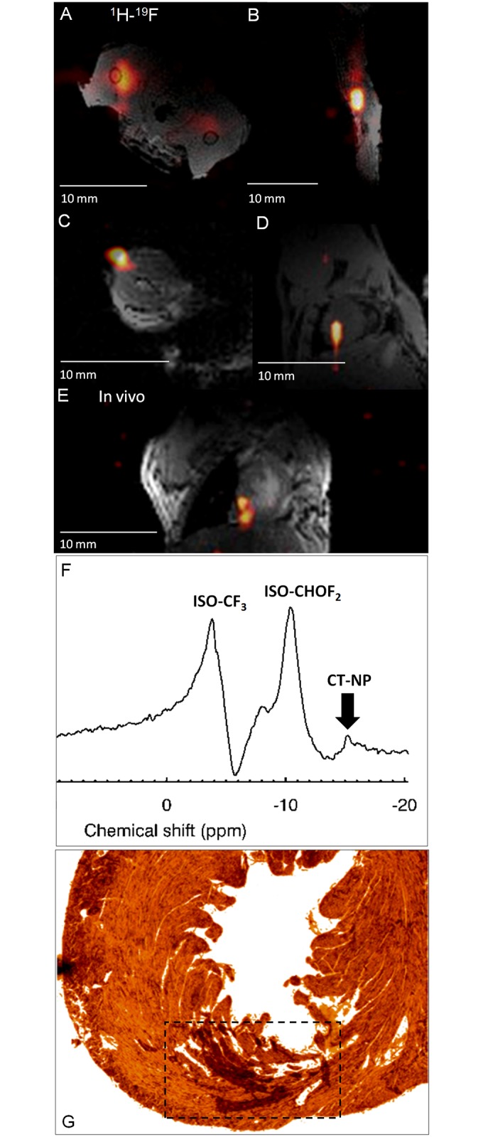
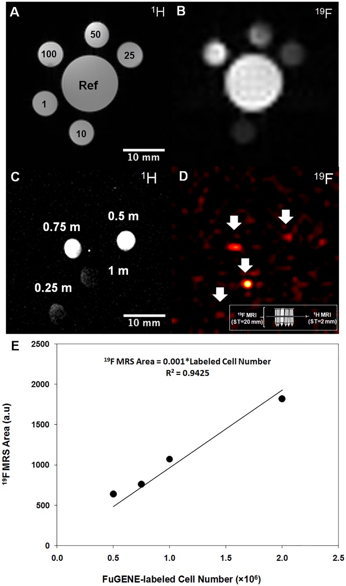
SPGR/SSFP proved favorable imaging sequences yielding good signal-to-noise ratio values. Confocal microscopy confirmed heterogeneity of cellular label uptake in CPCs. 19F MRI indicated lack of additional benefits upon label concentrations above 7.5-10 mg/ml/million cells. The minimum detectable CPC load was 500k (10k/voxel) in two-dimensional (2D) acquisitions (3-5 min) using the butterfly coil. Additionally, absolute 19F based concentration and intensity estimates (trifluoroacetic-acid solutions, macrophages, and labeled CPCs in vitro and post-CPC injections in the post-mortem state) scaled linearly with fluorine concentrations. Fast, quantitative cardiac 19F-MRI was demonstrated with SPGR/SSFP and MRS acquisitions spanning 3-5 min, using a butterfly coil.
The developed methodologies achieved in vivo cardiac 19F of exogenously injected labeled CPCs for the first time, accelerating imaging to a total acquisition of a few minutes, providing evidence for their potential for possible translational work.
a)实现对全氟冠醚(PFCE)标记的心脏祖干细胞(CPC)和骨源骨髓巨噬细胞进行心脏19F磁共振成像(MRI);b)确定标记浓度和细胞负载极限;c)实现基于光谱和图像的定量分析。
使用9.4 T的19F MRI/磁共振波谱(MRS)进行了扰相梯度回波(SPGR)、弛豫增强快速采集(RARE)和自由进动稳态(SSFP)脉冲序列的理论模拟和实验比较,以及体模验证。使用流式细胞术和共聚焦显微镜确认细胞标记成功。对于CPC和巨噬细胞浓度定量,使用转染试剂FuGENE进行体外和死后心脏验证。在小鼠心脏活体采集以及死后小鼠骨骼和心脏应用中证明了快速成像的可行性。
SPGR/SSFP被证明是产生良好信噪比的有利成像序列。共聚焦显微镜证实了CPC中细胞标记摄取的异质性。19F MRI表明,标记浓度高于7.5 - 10 mg/ml/百万细胞时没有额外益处。使用蝶形线圈在二维(2D)采集(3 - 5分钟)中,最小可检测CPC负载约为500k(~10k/体素)。此外,基于19F的绝对浓度和强度估计(三氟乙酸溶液、巨噬细胞以及体外和死后注射CPC后的标记CPC)与氟浓度呈线性比例关系。使用蝶形线圈,通过SPGR/SSFP和MRS采集,在3 - 5分钟内实现了快速、定量的心脏19F - MRI。
所开发的方法首次实现了对外源性注射标记的CPC进行体内心脏19F成像,将成像加速至总共几分钟的采集时间,为其可能的转化工作潜力提供了证据。