• 文献检索
  • 文档翻译
  • 深度研究
  • 学术资讯
  • Suppr Zotero 插件Zotero 插件
  • 邀请有礼
  • 套餐&价格
  • 历史记录
应用&插件
Suppr Zotero 插件Zotero 插件浏览器插件Mac 客户端Windows 客户端微信小程序
定价
高级版会员购买积分包购买API积分包
服务
文献检索文档翻译深度研究API 文档MCP 服务
关于我们
关于 Suppr公司介绍联系我们用户协议隐私条款
关注我们

Suppr 超能文献

核心技术专利:CN118964589B侵权必究
粤ICP备2023148730 号-1Suppr @ 2026

文献检索

告别复杂PubMed语法,用中文像聊天一样搜索,搜遍4000万医学文献。AI智能推荐,让科研检索更轻松。

立即免费搜索

文件翻译

保留排版,准确专业,支持PDF/Word/PPT等文件格式,支持 12+语言互译。

免费翻译文档

深度研究

AI帮你快速写综述,25分钟生成高质量综述,智能提取关键信息,辅助科研写作。

立即免费体验

NODDI-DTI:从健康白质中的扩散张量估计神经突方向和弥散参数。

NODDI-DTI: Estimating Neurite Orientation and Dispersion Parameters from a Diffusion Tensor in Healthy White Matter.

作者信息

Edwards Luke J, Pine Kerrin J, Ellerbrock Isabel, Weiskopf Nikolaus, Mohammadi Siawoosh

机构信息

Department of Neurophysics, Max Planck Institute for Human Cognitive and Brain Sciences, Leipzig, Germany.

Wellcome Trust Centre for Neuroimaging, UCL Institute of Neurology, University College London, London, United Kingdom.

出版信息

Front Neurosci. 2017 Dec 20;11:720. doi: 10.3389/fnins.2017.00720. eCollection 2017.

DOI:10.3389/fnins.2017.00720
PMID:29326546
原文链接:https://pmc.ncbi.nlm.nih.gov/articles/PMC5742359/
Abstract

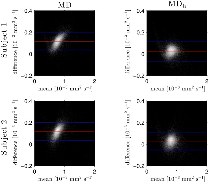
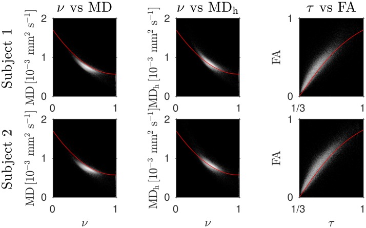
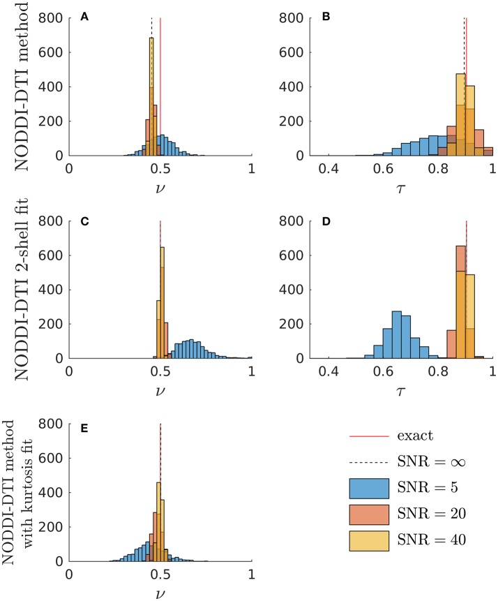
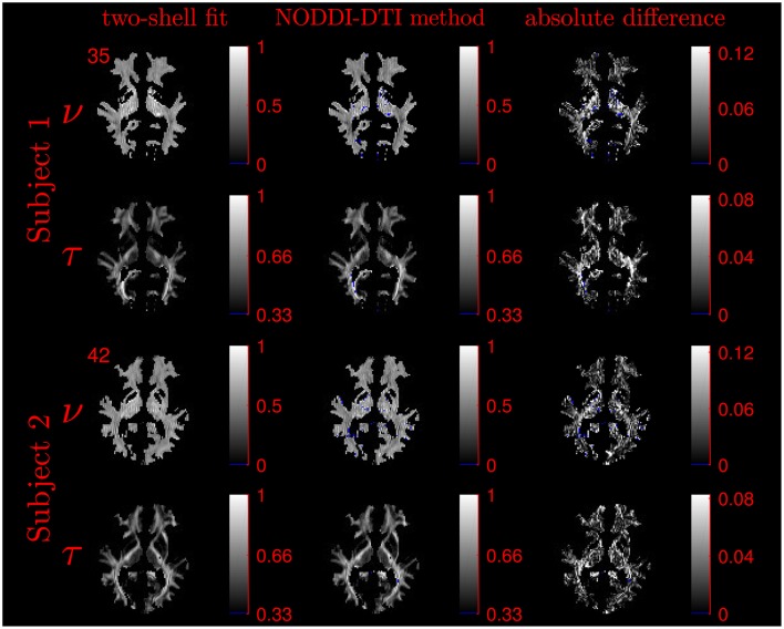
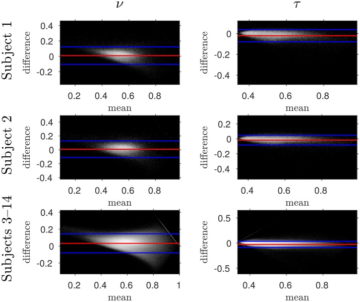
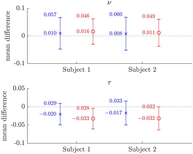
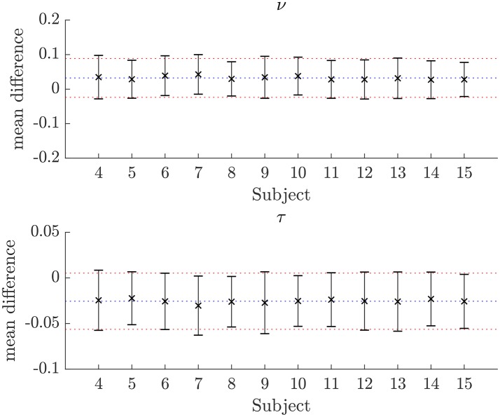
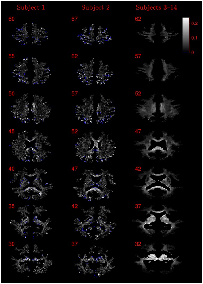
The NODDI-DTI signal model is a modification of the NODDI signal model that formally allows interpretation of standard single-shell DTI data in terms of biophysical parameters in healthy human white matter (WM). The NODDI-DTI signal model contains no CSF compartment, restricting application to voxels without CSF partial-volume contamination. This modification allowed derivation of analytical relations between parameters representing axon density and dispersion, and DTI invariants (MD and FA) from the NODDI-DTI signal model. These relations formally allow extraction of biophysical parameters from DTI data. NODDI-DTI parameters were estimated by applying the proposed analytical relations to DTI parameters estimated from the first shell of data, and compared to parameters estimated by fitting the NODDI-DTI model to both shells of data (reference dataset) in the WM of 14 diffusion datasets recorded with two different protocols, and in simulated data. The first two datasets were also fit to the NODDI-DTI model using only the first shell (as for DTI) of data. NODDI-DTI parameters estimated from DTI, and NODDI-DTI parameters estimated by fitting the model to the first shell of data gave similar errors compared to two-shell NODDI-DTI estimates. The simulations showed the NODDI-DTI method to be more noise-robust than the two-shell fitting procedure. The NODDI-DTI method gave unphysical parameter estimates in a small percentage of voxels, reflecting voxelwise DTI estimation error or NODDI-DTI model invalidity. In the course of evaluating the NODDI-DTI model, it was found that diffusional kurtosis strongly biased DTI-based MD values, and so, making assumptions based on healthy WM, a novel heuristic correction requiring only DTI data was derived and used to mitigate this bias. Since validations were only performed on healthy WM, application to grey matter or pathological WM would require further validation. Our results demonstrate NODDI-DTI to be a promising model and technique to interpret restricted datasets acquired for DTI analysis in healthy white matter with greater biophysical specificity, though its limitations must be borne in mind.

摘要

NODDI-DTI信号模型是对NODDI信号模型的一种修改,它正式允许根据健康人类白质(WM)中的生物物理参数来解释标准单壳DTI数据。NODDI-DTI信号模型不包含脑脊液(CSF)成分,因此仅限于应用于没有CSF部分容积污染的体素。这种修改使得能够从NODDI-DTI信号模型中推导出表示轴突密度和离散度的参数与DTI不变量(平均扩散率[MD]和各向异性分数[FA])之间的解析关系。这些关系正式允许从DTI数据中提取生物物理参数。通过将所提出的解析关系应用于从第一组数据估计的DTI参数来估计NODDI-DTI参数,并将其与通过将NODDI-DTI模型拟合到两组数据(参考数据集)而估计的参数进行比较,这些数据来自使用两种不同协议记录的14个扩散数据集中的WM以及模拟数据。前两个数据集也仅使用数据的第一组(如同DTI那样)拟合到NODDI-DTI模型。与双壳NODDI-DTI估计相比,从DTI估计的NODDI-DTI参数以及通过将模型拟合到第一组数据估计的NODDI-DTI参数具有相似的误差。模拟表明,NODDI-DTI方法比双壳拟合程序对噪声更具鲁棒性。NODDI-DTI方法在一小部分体素中给出了不符合物理实际的参数估计,这反映了体素级DTI估计误差或NODDI-DTI模型无效。在评估NODDI-DTI模型的过程中,发现扩散峰度强烈影响基于DTI的MD值,因此,基于健康WM做出假设,推导并使用了一种仅需要DTI数据的新型启发式校正方法来减轻这种偏差。由于验证仅在健康WM上进行,因此应用于灰质或病理性WM需要进一步验证。我们的结果表明,NODDI-DTI是一个有前景的模型和技术,能够以更高的生物物理特异性解释为健康白质DTI分析获取的受限数据集,不过必须牢记其局限性。

https://cdn.ncbi.nlm.nih.gov/pmc/blobs/9242/5742359/94998351b7c1/fnins-11-00720-g0009.jpg
https://cdn.ncbi.nlm.nih.gov/pmc/blobs/9242/5742359/e4918d6431ce/fnins-11-00720-g0001.jpg
https://cdn.ncbi.nlm.nih.gov/pmc/blobs/9242/5742359/6e091f03b0fb/fnins-11-00720-g0002.jpg
https://cdn.ncbi.nlm.nih.gov/pmc/blobs/9242/5742359/420144590956/fnins-11-00720-g0003.jpg
https://cdn.ncbi.nlm.nih.gov/pmc/blobs/9242/5742359/3dbb552ccd3c/fnins-11-00720-g0004.jpg
https://cdn.ncbi.nlm.nih.gov/pmc/blobs/9242/5742359/c5cee83f6541/fnins-11-00720-g0005.jpg
https://cdn.ncbi.nlm.nih.gov/pmc/blobs/9242/5742359/22d83d856d76/fnins-11-00720-g0006.jpg
https://cdn.ncbi.nlm.nih.gov/pmc/blobs/9242/5742359/c3ec70a8db41/fnins-11-00720-g0007.jpg
https://cdn.ncbi.nlm.nih.gov/pmc/blobs/9242/5742359/0894931c2318/fnins-11-00720-g0008.jpg
https://cdn.ncbi.nlm.nih.gov/pmc/blobs/9242/5742359/94998351b7c1/fnins-11-00720-g0009.jpg

相似文献

1
NODDI-DTI: Estimating Neurite Orientation and Dispersion Parameters from a Diffusion Tensor in Healthy White Matter.NODDI-DTI:从健康白质中的扩散张量估计神经突方向和弥散参数。
Front Neurosci. 2017 Dec 20;11:720. doi: 10.3389/fnins.2017.00720. eCollection 2017.
2
Assessing Microstructural Substrates of White Matter Abnormalities: A Comparative Study Using DTI and NODDI.评估白质异常的微观结构基础:一项使用扩散张量成像(DTI)和神经突方向离散与密度成像(NODDI)的比较研究
PLoS One. 2016 Dec 21;11(12):e0167884. doi: 10.1371/journal.pone.0167884. eCollection 2016.
3
Neurite orientation dispersion and density imaging in myelin oligodendrocyte glycoprotein antibody-associated disease and neuromyelitis optica spectrum disorders.髓鞘少突胶质细胞糖蛋白抗体相关疾病和视神经脊髓炎谱系障碍中的神经突方向离散度与密度成像
Mult Scler Relat Disord. 2025 Mar;95:106324. doi: 10.1016/j.msard.2025.106324. Epub 2025 Feb 8.
4
Differences in Gaussian diffusion tensor imaging and non-Gaussian diffusion kurtosis imaging model-based estimates of diffusion tensor invariants in the human brain.基于高斯扩散张量成像和非高斯扩散峰度成像模型的人脑扩散张量不变量估计差异。
Med Phys. 2016 May;43(5):2464. doi: 10.1118/1.4946819.
5
Differentiation of Edematous, Tumoral and Normal Areas of Brain Using Diffusion Tensor and Neurite Orientation Dispersion and Density Imaging.利用扩散张量及神经突方向离散度与密度成像对脑内水肿、肿瘤及正常区域进行鉴别
J Biomed Phys Eng. 2018 Sep 1;8(3):251-260. eCollection 2018 Sep.
6
A systematic review of microstructural abnormalities in multiple sclerosis detected with NODDI and DTI models of diffusion-weighted magnetic resonance imaging.基于磁共振弥散加权成像 NODDI 和 DTI 模型检测多发性硬化的微观结构异常的系统评价。
Magn Reson Imaging. 2023 Dec;104:61-71. doi: 10.1016/j.mri.2023.09.010. Epub 2023 Sep 27.
7
NODDI reproducibility and variability with magnetic field strength: A comparison between 1.5 T and 3 T.NODDI在不同磁场强度下的可重复性和变异性:1.5T与3T的比较。
Hum Brain Mapp. 2016 Dec;37(12):4550-4565. doi: 10.1002/hbm.23328. Epub 2016 Aug 1.
8
Neurite orientation dispersion and density imaging of the healthy cervical spinal cord in vivo.健康颈椎脊髓的神经纤维取向分散和密度成像。
Neuroimage. 2015 May 1;111:590-601. doi: 10.1016/j.neuroimage.2015.01.045. Epub 2015 Jan 31.
9
Diffusion Tensor Model links to Neurite Orientation Dispersion and Density Imaging at high b-value in Cerebral Cortical Gray Matter.弥散张量模型与大脑皮质灰质的高 b 值下神经纤维取向弥散和密度成像相关联。
Sci Rep. 2019 Aug 22;9(1):12246. doi: 10.1038/s41598-019-48671-7.
10
White Matter Microstructural Alterations in Type 2 Diabetes: A Combined UK Biobank Study of Diffusion Tensor Imaging and Neurite Orientation Dispersion and Density Imaging.2型糖尿病中的白质微观结构改变:英国生物银行扩散张量成像与神经突方向离散度和密度成像的联合研究
Medicina (Kaunas). 2025 Mar 6;61(3):455. doi: 10.3390/medicina61030455.

引用本文的文献

1
ACID: A comprehensive toolbox for image processing and modeling of brain, spinal cord, and ex vivo diffusion MRI data.ACID:用于脑、脊髓及离体扩散磁共振成像数据的图像处理和建模的综合工具箱。
Imaging Neurosci (Camb). 2024 Sep 13;2. doi: 10.1162/imag_a_00288. eCollection 2024.
2
The trouble with free-water elimination using single-shell diffusion MRI data: A case study in ageing.使用单壳扩散磁共振成像数据进行自由水消除的问题:一项关于衰老的案例研究。
Imaging Neurosci (Camb). 2024 Aug 1;2. doi: 10.1162/imag_a_00252. eCollection 2024.
3
White matter characterization in regions of edema surrounding meningioma brain tumor using diffusion MRI: A comparative study of DTI and NODDI.

本文引用的文献

1
Brain microstructure by multi-modal MRI: Is the whole greater than the sum of its parts?多模态 MRI 下的脑微观结构:整体是否大于其各部分之和?
Neuroimage. 2018 Nov 15;182:117-127. doi: 10.1016/j.neuroimage.2017.10.052. Epub 2017 Nov 4.
2
Four in vivo g-ratio-weighted imaging methods: Comparability and repeatability at the group level.四种活体 g 比值加权成像方法:在组水平上的可比性和可重复性。
Hum Brain Mapp. 2018 Jan;39(1):24-41. doi: 10.1002/hbm.23858. Epub 2017 Nov 1.
3
Promise and pitfalls of g-ratio estimation with MRI.MRI 估计 g-比值的前景与陷阱。
使用扩散磁共振成像对脑膜瘤脑肿瘤周围水肿区域的白质特征进行研究:扩散张量成像(DTI)与神经突方向离散与密度成像(NODDI)的对比研究
medRxiv. 2025 Apr 8:2025.04.07.25325393. doi: 10.1101/2025.04.07.25325393.
4
Altered white matter microstructural integrity in patients with postherpetic neuralgia: a combined DTI and DTI-NODDI study.带状疱疹后神经痛患者白质微结构完整性改变:一项联合扩散张量成像(DTI)和扩散张量成像-神经突方向离散与密度成像(DTI-NODDI)的研究
Front Neurosci. 2025 Feb 18;19:1552961. doi: 10.3389/fnins.2025.1552961. eCollection 2025.
5
Temporal dynamics of white and gray matter plasticity during motor skill acquisition: a comparative diffusion tensor imaging and multiparametric mapping analysis.运动技能习得过程中白质和灰质可塑性的时间动态:扩散张量成像和多参数映射分析的比较。
Cereb Cortex. 2024 Aug 1;34(8). doi: 10.1093/cercor/bhae344.
6
Microstructure predicts non-motor outcomes following deep brain stimulation in Parkinson's disease.微观结构可预测帕金森病患者深部脑刺激后的非运动结果。
NPJ Parkinsons Dis. 2024 May 18;10(1):104. doi: 10.1038/s41531-024-00717-y.
7
Brain temperature and free water increases after mild COVID-19 infection.轻度 COVID-19 感染后大脑温度和自由水增加。
Sci Rep. 2024 Mar 28;14(1):7450. doi: 10.1038/s41598-024-57561-6.
8
Neural Correlates of Obesity and Inflammation in Children and Adolescents with Congenital Adrenal Hyperplasia.先天性肾上腺皮质增生症儿童及青少年肥胖与炎症的神经关联
Horm Res Paediatr. 2025;98(2):174-184. doi: 10.1159/000537847. Epub 2024 Feb 19.
9
In Vivo Characterization of Cerebellar Peduncles in Chronic Ankle Instability: A Single and Multishell Diffusion-Weighted Imaging Study.慢性踝关节不稳患者小脑脚的体内特征:一项单壳层和多壳层弥散加权成像研究。
Sports Health. 2024 Jan-Feb;16(1):38-46. doi: 10.1177/19417381231156544. Epub 2023 Mar 2.
10
Insights and improvements in correspondence between axonal volume fraction measured with diffusion-weighted MRI and electron microscopy.弥散加权 MRI 测量的轴突体积分数与电子显微镜之间相关性的见解和改进。
NMR Biomed. 2024 Mar;37(3):e5070. doi: 10.1002/nbm.5070. Epub 2023 Dec 14.
Neuroimage. 2018 Nov 15;182:80-96. doi: 10.1016/j.neuroimage.2017.08.038. Epub 2017 Aug 16.
4
Image quality transfer and applications in diffusion MRI.扩散磁共振成像中的图像质量传递及其应用
Neuroimage. 2017 May 15;152:283-298. doi: 10.1016/j.neuroimage.2017.02.089. Epub 2017 Mar 3.
5
Challenges and opportunities for brainstem neuroimaging with ultrahigh field MRI.超高场 MRI 用于脑干神经影像学的挑战与机遇。
Neuroimage. 2018 Mar;168:412-426. doi: 10.1016/j.neuroimage.2017.02.052. Epub 2017 Feb 21.
6
Neurite density imaging versus imaging of microscopic anisotropy in diffusion MRI: A model comparison using spherical tensor encoding.扩散磁共振成像中神经突密度成像与微观各向异性成像的比较:使用球张量编码的模型对比
Neuroimage. 2017 Feb 15;147:517-531. doi: 10.1016/j.neuroimage.2016.11.053. Epub 2016 Nov 27.
7
Fast imaging of mean, axial and radial diffusion kurtosis.平均、轴向和径向扩散峰度的快速成像。
Neuroimage. 2016 Nov 15;142:381-393. doi: 10.1016/j.neuroimage.2016.08.022. Epub 2016 Aug 15.
8
Multi-compartment microscopic diffusion imaging.多室微观扩散成像
Neuroimage. 2016 Oct 1;139:346-359. doi: 10.1016/j.neuroimage.2016.06.002. Epub 2016 Jun 6.
9
NODDI and Tensor-Based Microstructural Indices as Predictors of Functional Connectivity.基于体素的神经突方向离散度与密度成像(NODDI)和基于张量的微观结构指数作为功能连接性的预测指标
PLoS One. 2016 Apr 14;11(4):e0153404. doi: 10.1371/journal.pone.0153404. eCollection 2016.
10
A prospective microstructure imaging study in mixed-martial artists using geometric measures and diffusion tensor imaging: methods and findings.一项使用几何测量和扩散张量成像技术对综合格斗运动员进行的前瞻性微观结构成像研究:方法与发现。
Brain Imaging Behav. 2017 Jun;11(3):698-711. doi: 10.1007/s11682-016-9546-1.