• 文献检索
  • 文档翻译
  • 深度研究
  • 学术资讯
  • Suppr Zotero 插件Zotero 插件
  • 邀请有礼
  • 套餐&价格
  • 历史记录
应用&插件
Suppr Zotero 插件Zotero 插件浏览器插件Mac 客户端Windows 客户端微信小程序
定价
高级版会员购买积分包购买API积分包
服务
文献检索文档翻译深度研究API 文档MCP 服务
关于我们
关于 Suppr公司介绍联系我们用户协议隐私条款
关注我们

Suppr 超能文献

核心技术专利:CN118964589B侵权必究
粤ICP备2023148730 号-1Suppr @ 2026

文献检索

告别复杂PubMed语法,用中文像聊天一样搜索,搜遍4000万医学文献。AI智能推荐,让科研检索更轻松。

立即免费搜索

文件翻译

保留排版,准确专业,支持PDF/Word/PPT等文件格式,支持 12+语言互译。

免费翻译文档

深度研究

AI帮你快速写综述,25分钟生成高质量综述,智能提取关键信息,辅助科研写作。

立即免费体验

中央静脉征可将多发性硬化与中枢神经系统炎症性血管病区分开来。

Central vein sign differentiates Multiple Sclerosis from central nervous system inflammatory vasculopathies.

机构信息

Department of Neurology, Université Libre de Bruxelles, Brussels, Belgium.

Department of Neurology, Brugmann University Hospital, Université Libre de Bruxelles, Brussels, Belgium.

出版信息

Ann Neurol. 2018 Feb;83(2):283-294. doi: 10.1002/ana.25146. Epub 2018 Feb 15.

DOI:10.1002/ana.25146
PMID:29328521
原文链接:https://pmc.ncbi.nlm.nih.gov/articles/PMC5901412/
Abstract

OBJECTIVES

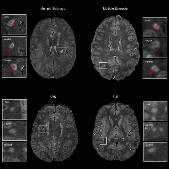
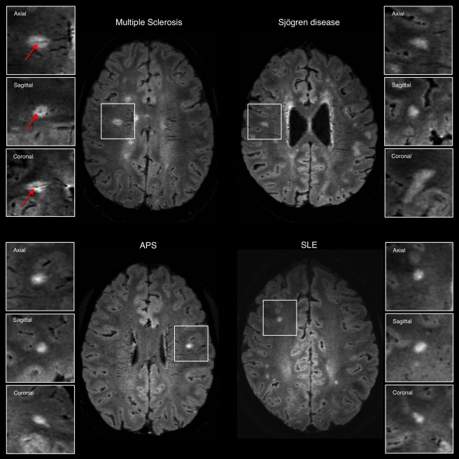
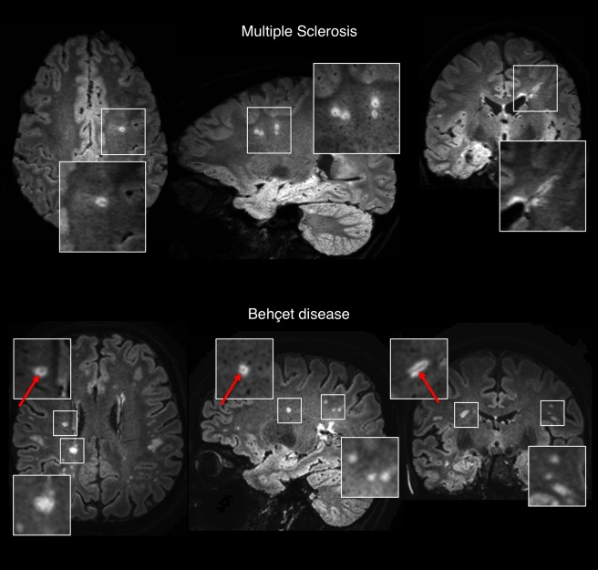
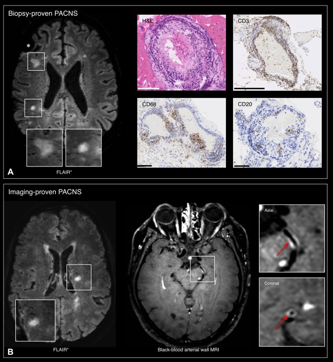
In multiple sclerosis (MS), magnetic resonance imaging (MRI) is a sensitive tool for detecting white matter lesions, but its diagnostic specificity is still suboptimal; ambiguous cases are frequent in clinical practice. Detection of perivenular lesions in the brain (the "central vein sign") improves the pathological specificity of MS diagnosis, but comprehensive evaluation of this MRI biomarker in MS-mimicking inflammatory and/or autoimmune diseases, such as central nervous system (CNS) inflammatory vasculopathies, is lacking. In a multicenter study, we assessed the frequency of perivenular lesions in MS versus systemic autoimmune diseases with CNS involvement and primary angiitis of the CNS (PACNS).

METHODS

In 31 patients with inflammatory CNS vasculopathies and 52 with relapsing-remitting MS, 3-dimensional T2*-weighted and T2-fluid-attenuated inversion recovery images were obtained during a single MRI acquisition after gadolinium injection. For each lesion, the central vein sign was evaluated according to consensus guidelines. For each patient, lesion count, volume, and brain location, as well as fulfillment of dissemination in space MRI criteria, were assessed.

RESULTS

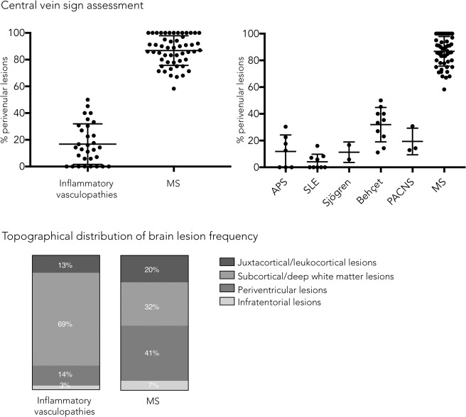
MS showed higher frequency of perivenular lesions (median = 88%) than did inflammatory CNS vasculopathies (14%), without overlap between groups or differences between 3T and 1.5T MRI. Among inflammatory vasculopathies, Behçet disease showed the highest median frequency of perivenular lesions (34%), followed by PACNS (14%), antiphospholipid syndromes (12%), Sjögren syndrome (11%), and systemic lupus erythematosus (0%). When a threshold of 50% perivenular lesions was applied, central vein sign discriminated MS from inflammatory vasculopathies with a diagnostic accuracy of 100%.

INTERPRETATION

The central vein sign differentiates inflammatory CNS vasculopathies from MS at standard clinical magnetic field strengths. Ann Neurol 2018;83:283-294.

摘要

目的

在多发性硬化症(MS)中,磁共振成像(MRI)是检测脑白质病变的敏感工具,但诊断特异性仍不理想;在临床实践中,存在大量模棱两可的病例。检测脑内血管周围病变(“中央静脉征”)可提高 MS 诊断的病理特异性,但对 MS 样炎症和/或自身免疫性疾病(如中枢神经系统(CNS)炎症性血管病)中这种 MRI 生物标志物的综合评估尚缺乏。在一项多中心研究中,我们评估了 MS 与伴 CNS 受累的系统性自身免疫性疾病及原发性中枢神经系统血管炎(PACNS)中静脉周围病变的发生率。

方法

在 31 例炎症性 CNS 血管病患者和 52 例复发缓解型 MS 患者中,在单次 MRI 采集后静脉注射钆剂,获得三维 T2*-加权和 T2 液体衰减反转恢复图像。根据共识指南评估每个病变的中央静脉征。评估每个患者的病变计数、体积和脑区位置,以及是否符合空间弥散 MRI 标准。

结果

MS 静脉周围病变的频率(中位数=88%)高于炎症性 CNS 血管病(14%),两组间无重叠,3T 和 1.5T MRI 之间也无差异。在炎症性血管病中,贝赫切特病静脉周围病变的频率最高(中位数=34%),其次是 PACNS(中位数=14%)、抗磷脂综合征(中位数=12%)、干燥综合征(中位数=11%)和系统性红斑狼疮(中位数=0%)。当静脉周围病变 50%作为阈值时,中央静脉征可以 100%的准确率将 MS 与炎症性血管病区分开来。

结论

在标准临床磁场强度下,中央静脉征可区分炎症性 CNS 血管病与 MS。

https://cdn.ncbi.nlm.nih.gov/pmc/blobs/3a09/5901412/e22c919feafd/ANA-83-283-g005.jpg
https://cdn.ncbi.nlm.nih.gov/pmc/blobs/3a09/5901412/bdc4e90b6c2a/ANA-83-283-g001.jpg
https://cdn.ncbi.nlm.nih.gov/pmc/blobs/3a09/5901412/44f202c77e7c/ANA-83-283-g002.jpg
https://cdn.ncbi.nlm.nih.gov/pmc/blobs/3a09/5901412/7a4facd05bf5/ANA-83-283-g003.jpg
https://cdn.ncbi.nlm.nih.gov/pmc/blobs/3a09/5901412/cf523fa4196a/ANA-83-283-g004.jpg
https://cdn.ncbi.nlm.nih.gov/pmc/blobs/3a09/5901412/e22c919feafd/ANA-83-283-g005.jpg

相似文献

1
Central vein sign differentiates Multiple Sclerosis from central nervous system inflammatory vasculopathies.中央静脉征可将多发性硬化与中枢神经系统炎症性血管病区分开来。
Ann Neurol. 2018 Feb;83(2):283-294. doi: 10.1002/ana.25146. Epub 2018 Feb 15.
2
Evaluation of the Central Vein Sign as a Diagnostic Imaging Biomarker in Multiple Sclerosis.中央静脉征作为多发性硬化症诊断影像学生物标志物的评估。
JAMA Neurol. 2019 Dec 1;76(12):1446-1456. doi: 10.1001/jamaneurol.2019.2478.
3
Distinct regional brain atrophy pattern in multiple sclerosis and neuropsychiatric systemic lupus erythematosus patients.多发性硬化症和神经精神性系统性红斑狼疮患者不同的脑区萎缩模式。
Lupus. 2018 Sep;27(10):1624-1635. doi: 10.1177/0961203318781004. Epub 2018 Jun 27.
4
Limited utility of current MRI criteria for distinguishing multiple sclerosis from common mimickers: primary and secondary CNS vasculitis, lupus and Sjogren's syndrome.当前 MRI 标准在鉴别多发性硬化与常见类似疾病(原发性和继发性中枢神经系统血管炎、狼疮和干燥综合征)方面的应用有限。
Mult Scler. 2014 Jan;20(1):57-63. doi: 10.1177/1352458513491329. Epub 2013 Jun 4.
5
Multiple sclerosis: High prevalence of the 'central vein' sign in white matter lesions on susceptibility-weighted images.多发性硬化症:磁敏感加权成像上白质病变中“中央静脉”征的高发生率。
Neuroradiol J. 2018 Aug;31(4):356-361. doi: 10.1177/1971400918763577. Epub 2018 Mar 22.
6
Comparison of Multiple Sclerosis Cortical Lesion Types Detected by Multicontrast 3T and 7T MRI.多对比度 3T 和 7T MRI 检测多发性硬化皮质病变类型的比较。
AJNR Am J Neuroradiol. 2019 Jul;40(7):1162-1169. doi: 10.3174/ajnr.A6099. Epub 2019 Jun 20.
7
Susceptibility-weighted imaging in the differential diagnosis of autoimmune central nervous system vasculitis and multiple sclerosis.磁敏感加权成像在自身免疫性中枢神经系统血管炎和多发性硬化鉴别诊断中的应用。
Mult Scler Relat Disord. 2019 Aug;33:70-74. doi: 10.1016/j.msard.2019.05.012. Epub 2019 May 23.
8
7T MRI cerebral leptomeningeal enhancement is common in relapsing-remitting multiple sclerosis and is associated with cortical and thalamic lesions.7T MRI 脑软脑膜强化在复发缓解型多发性硬化症中很常见,与皮质和丘脑病变有关。
Mult Scler. 2020 Feb;26(2):177-187. doi: 10.1177/1352458519885106. Epub 2019 Nov 12.
9
The "central vein sign" in patients with diagnostic "red flags" for multiple sclerosis: A prospective multicenter 3T study.多发性硬化症诊断“红旗”患者的“中央静脉征”:一项前瞻性多中心3T研究。
Mult Scler. 2020 Apr;26(4):421-432. doi: 10.1177/1352458519876031. Epub 2019 Sep 19.
10
Morphology-Specific Discrimination between MS White Matter Lesions and Benign White Matter Hyperintensities Using Ultra-High-Field MRI.超高场 MRI 对 MS 脑白质病变与良性脑白质高信号的形态学鉴别诊断
AJNR Am J Neuroradiol. 2018 Aug;39(8):1473-1479. doi: 10.3174/ajnr.A5705. Epub 2018 Jun 21.

引用本文的文献

1
The Cerebral Lymphatic System: Function, Controversies, and Therapeutic Approaches for Central Nervous System Diseases.脑淋巴系统:中枢神经系统疾病的功能、争议及治疗方法
Cell Mol Neurobiol. 2025 Aug 19;45(1):80. doi: 10.1007/s10571-025-01598-2.
2
Widespread venous thrombosis: Unveiling a complex case of Behçet's disease with a literature perspective.广泛静脉血栓形成:从文献角度揭示一例复杂的白塞病病例
Open Med (Wars). 2025 Aug 6;20(1):20251199. doi: 10.1515/med-2025-1199. eCollection 2025.
3
A pilot study assessing the clinical utility of deep learning-reconstructed 3D-echo-planar-imaging-based quantitative susceptibility mapping in multiple sclerosis.

本文引用的文献

1
Diagnostic performance of central vein sign for multiple sclerosis with a simplified three-lesion algorithm.简化三病灶算法下中央静脉征对多发性硬化症的诊断性能。
Mult Scler. 2018 May;24(6):750-757. doi: 10.1177/1352458517726383. Epub 2017 Aug 18.
2
The tension between early diagnosis and misdiagnosis of multiple sclerosis.多发性硬化症早期诊断与误诊之间的矛盾关系。
Nat Rev Neurol. 2017 Sep;13(9):567-572. doi: 10.1038/nrneurol.2017.106. Epub 2017 Aug 11.
3
Myelopathy in Behçet's disease: The Bagel Sign.贝切特病性脊髓病:“贝果征”。
一项评估基于深度学习重建的三维回波平面成像定量磁化率图谱在多发性硬化症中的临床应用价值的试点研究。
Front Neurosci. 2025 Jul 16;19:1544376. doi: 10.3389/fnins.2025.1544376. eCollection 2025.
4
Association of a giant developmental venous anomaly and acute disseminated encephalomyelitis: A case report and magnetic resonance perfusion study.巨大发育性静脉异常与急性播散性脑脊髓炎的关联:一例报告及磁共振灌注研究
Neuroradiol J. 2025 Jun 30:19714009251356989. doi: 10.1177/19714009251356989.
5
Diagnostic Accuracy of Isotropic FLAIR-T2* Fusion Imaging for Central Vein Sign Detection in Multiple Sclerosis: a Comparative Study at 1.5 T and 3 T.各向同性液体衰减反转恢复序列与T2*融合成像检测多发性硬化症中央静脉征的诊断准确性:1.5T和3T的对比研究
Clin Neuroradiol. 2025 Jun 16. doi: 10.1007/s00062-025-01531-6.
6
Clinical integration of brain and cord MRI features improves differential diagnosis of multiple sclerosis.脑和脊髓MRI特征的临床整合改善了多发性硬化的鉴别诊断。
J Neurol. 2025 May 11;272(6):388. doi: 10.1007/s00415-025-13124-x.
7
The Kappa Free Light Chains Index and Central Vein Sign: Two New Biomarkers for Multiple Sclerosis Diagnosis.游离κ轻链指数与中央静脉征:多发性硬化诊断的两种新生物标志物。
Neurol Ther. 2025 Jun;14(3):711-731. doi: 10.1007/s40120-025-00737-7. Epub 2025 Apr 6.
8
Post-contrast Susceptibility Weighted Imaging in Multiple Sclerosis MRI Improves the Detection of Enhancing Lesions.多发性硬化症磁共振成像中的对比剂后 susceptibility 加权成像可提高强化病灶的检测率。
Clin Neuroradiol. 2025 Mar 7. doi: 10.1007/s00062-025-01508-5.
9
Multiple sclerosis diagnosis and its differential diagnosis in patients presenting with type four 'mirror pattern' CSF oligoclonal bands.出现4型“镜像模式”脑脊液寡克隆带患者的多发性硬化症诊断及其鉴别诊断
J Neurol. 2025 Feb 15;272(3):207. doi: 10.1007/s00415-025-12947-y.
10
Incorporation of the central vein sign into the McDonald criteria.将中央静脉征纳入麦克唐纳标准。
Mult Scler Relat Disord. 2025 Jan;93:106182. doi: 10.1016/j.msard.2024.106182. Epub 2024 Nov 25.
Ann Neurol. 2017 Aug;82(2):288-298. doi: 10.1002/ana.25004. Epub 2017 Aug 10.
4
Subtypes of primary angiitis of the CNS identified by MRI patterns reflect the size of affected vessels.MRI 表现确定的原发性中枢神经系统血管炎亚型与受累血管大小相关。
J Neurol Neurosurg Psychiatry. 2017 Sep;88(9):749-755. doi: 10.1136/jnnp-2017-315691. Epub 2017 Jul 13.
5
FLAIR* to visualize veins in white matter lesions: A new tool for the diagnosis of multiple sclerosis?FLAIR 序列对脑白质病变内静脉的显示:多发性硬化症诊断的新工具?
Eur Radiol. 2017 Oct;27(10):4257-4263. doi: 10.1007/s00330-017-4822-z. Epub 2017 Apr 13.
6
Diagnosis of multiple sclerosis: progress and challenges.多发性硬化症的诊断:进展与挑战。
Lancet. 2017 Apr 1;389(10076):1336-1346. doi: 10.1016/S0140-6736(16)30959-X. Epub 2016 Nov 24.
7
The central vein sign and its clinical evaluation for the diagnosis of multiple sclerosis: a consensus statement from the North American Imaging in Multiple Sclerosis Cooperative.中央静脉征及其临床评估在多发性硬化诊断中的应用:北美多发性硬化影像学合作组的共识声明。
Nat Rev Neurol. 2016 Dec;12(12):714-722. doi: 10.1038/nrneurol.2016.166. Epub 2016 Nov 11.
8
2016 American College of Rheumatology/European League Against Rheumatism classification criteria for primary Sjögren's syndrome: A consensus and data-driven methodology involving three international patient cohorts.2016 年美国风湿病学会/欧洲抗风湿病联盟原发性干燥综合征分类标准:一项涉及三个国际患者队列的共识和数据驱动方法。
Ann Rheum Dis. 2017 Jan;76(1):9-16. doi: 10.1136/annrheumdis-2016-210571. Epub 2016 Oct 26.
9
"Central vessel sign" on 3T FLAIR* MRI for the differentiation of multiple sclerosis from migraine.3T FLAIR* MRI 上的“中央血管征”在鉴别多发性硬化与偏头痛中的作用。
Ann Clin Transl Neurol. 2015 Dec 16;3(2):82-7. doi: 10.1002/acn3.273. eCollection 2016 Feb.
10
MRI criteria for the diagnosis of multiple sclerosis: MAGNIMS consensus guidelines.多发性硬化诊断的MRI标准:MAGNIMS共识指南。
Lancet Neurol. 2016 Mar;15(3):292-303. doi: 10.1016/S1474-4422(15)00393-2. Epub 2016 Jan 26.