Maggi Pietro, Absinta Martina, Sati Pascal, Perrotta Gaetano, Massacesi Luca, Dachy Bernard, Pot Caroline, Meuli Reto, Reich Daniel S, Filippi Massimo, Pasquier Renaud Du, Théaudin Marie
Department of Neurology, Center of Clinical Neurosciences, Lausanne University Hospital, Lausanne, Switzerland/ Department of Neurology, Hôpital Erasme, Université Libre de Bruxelles, Brussels, Belgium.
Translational Neuroradiology Section, National Institute of Neurological Disorders and Stroke, National Institutes of Health, Bethesda, MD, USA/ Department of Neurology, San Raffaele Scientific Institute, Vita-Salute San Raffaele University, Milan, Italy/ Neuroimaging Research Unit, Institute of Experimental Neurology, Division of Neuroscience, San Raffaele Scientific Institute, Vita-Salute San Raffaele University, Milan, Italy.
Mult Scler. 2020 Apr;26(4):421-432. doi: 10.1177/1352458519876031. Epub 2019 Sep 19.
The central vein sign (CVS) has been shown to help in the differential diagnosis of multiple sclerosis (MS), but most prior studies are retrospective.
To prospectively assess the diagnostic predictive value of the CVS in diagnostically difficult cases.
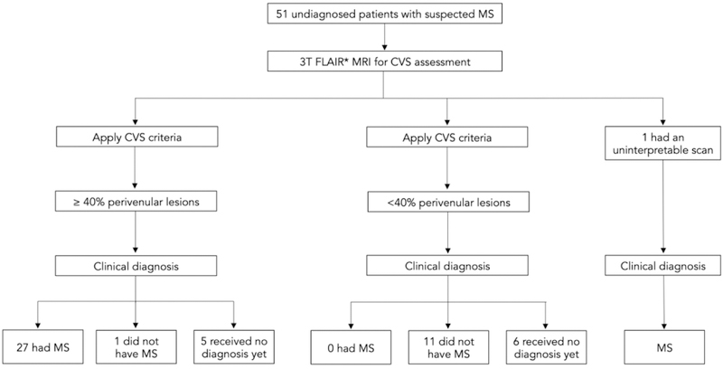
In this prospective multicenter study, 51 patients with suspected MS who had clinical, imaging, or laboratory "red flags" (i.e. features atypical for MS) underwent 3T fluid-attenuated inversion recovery (FLAIR*) magnetic resonance imaging (MRI) for CVS assessment. After the diagnostic work-up, expert clinicians blinded to the results of the CVS assessment came to a clinical diagnosis. The value of the CVS to prospectively predict an MS diagnosis was assessed.
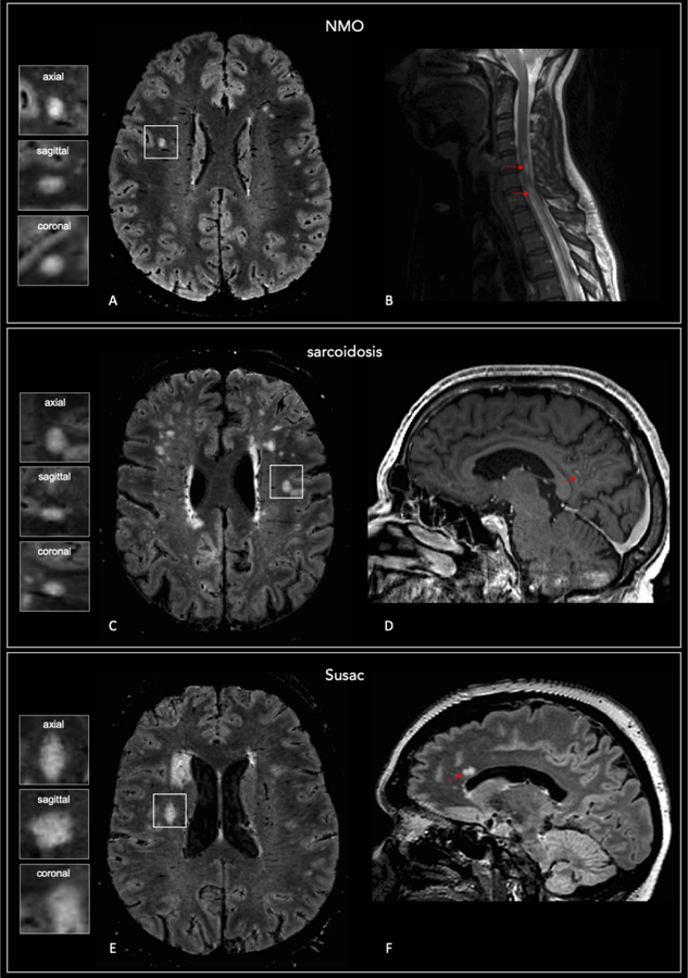
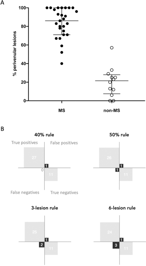
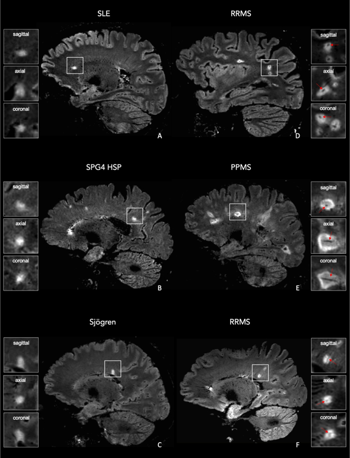
Of the 39 patients who received a clinical diagnosis by the end of the study, 27 had MS and 12 received a non-MS diagnosis that included systemic lupus erythematosus, sarcoidosis, migraine, Sjögren disease, SPG4-spastic-paraparesis, neuromyelitis optica, and Susac syndrome. The percentage of perivenular lesions was higher in MS (median = 86%) compared to non-MS (median = 21%; < 0.0001) patients. A 40% perivenular lesion cutoff was associated with 97% accuracy and a 96% positive/100% negative predictive value.
The CVS detected on 3T FLAIR* images can accurately predict an MS diagnosis in patients suspected to have MS, but with atypical clinical, laboratory, and imaging features.
中央静脉征(CVS)已被证明有助于多发性硬化症(MS)的鉴别诊断,但大多数先前的研究都是回顾性的。
前瞻性评估CVS在诊断困难病例中的诊断预测价值。
在这项前瞻性多中心研究中,51例疑似MS且有临床、影像或实验室“红旗”(即MS非典型特征)的患者接受了3T液体衰减反转恢复(FLAIR*)磁共振成像(MRI)以评估CVS。在诊断检查后,对CVS评估结果不知情的专家临床医生做出临床诊断。评估CVS前瞻性预测MS诊断的价值。
在研究结束时获得临床诊断的39例患者中,27例患有MS,12例获得非MS诊断,包括系统性红斑狼疮、结节病、偏头痛、干燥综合征、SPG4痉挛性截瘫、视神经脊髓炎和Susac综合征。与非MS患者(中位数 = 21%;<0.0001)相比,MS患者(中位数 = 86%)的血管周围病变百分比更高。血管周围病变截断值为40%时,准确率为97%,阳性预测值为96%,阴性预测值为100%。
在3T FLAIR*图像上检测到的CVS可以准确预测疑似MS但具有非典型临床、实验室和影像特征患者的MS诊断。