Tsutsui Yuji, Awamoto Shinichi, Himuro Kazuhiko, Umezu Yoshiyuki, Baba Shingo, Sasaki Masayuki
Division of Radiology, Department of Medical Technology, Kyushu University Hospital, Fukuoka, Japan.
Medical Quantum Science Course, Department of Health Sciences, Graduate School of Medical Sciences, Kyushu University, Fukuoka, Japan.
Asia Ocean J Nucl Med Biol. 2018 Winter;6(1):15-23. doi: 10.22038/aojnmb.2017.26684.1186.
The aim of this study is to examine the effect of different smoothing filters on the image quality and SUV to achieve the guideline recommended positron emission tomography (PET) image without harmonization.
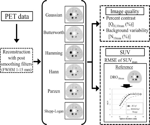
We used a Biograph mCT PET scanner. A National Electrical Manufacturers Association (NEMA) the International Electrotechnical Commission (IEC) body phantom was filled with F solution with a background activity of 2.65 kBq/mL and a sphere-to-background ratio of 4. PET images obtained with the Biograph mCT PET scanner were reconstructed using the ordered subsets-expectation maximization (OSEM) algorithm with time-of-flight (TOF) models (iteration, 2; subset, 21); smoothing filters including the Gaussian, Butterworth, Hamming, Hann, Parzen, and Shepp-Logan filters with various full width at half maximum (FWHM) values (1-15 mm) were applied. The image quality was physically assessed according to the percent contrast (Q), background variability (N), standardized uptake value (SUV), and recovery coefficient (RC). The results were compared with the guideline recommended range proposed by the Japanese Society of Nuclear Medicine and the Japanese Society of Nuclear Medicine Technology. The PET digital phantom was developed from the digital reference object (DRO) of the NEMA IEC body phantom smoothed using a Gaussian filter with a 10-mm FWHM and defined as the reference image. The difference in the SUV between the PET image and the reference image was evaluated according to the root mean squared error (RMSE).
The FWHMs of the Gaussian, Butterworth, Hamming, Hann, Parzen, and Shepp-Logan filters that satisfied the image quality of the FDG-PET/CT standardization guideline criteria were 8-12 mm, 9-11 mm, 9-13 mm, 10-13 mm, 9-11 mm, and 12-15 mm, respectively. The FWHMs of the Gaussian, Butterworth, Hamming, Hann, Parzen, and Shepp-Logan filters that provided the smallest RMSE between the PET images and the 3D digital phantom were 7 mm, 8 mm, 8 mm, 8 mm, 7 mm, and 11 mm, respectively.
The suitable FWHM for image quality or SUV depends on the type of smoothing filter that is applied.
本研究旨在探讨不同平滑滤波器对图像质量和标准化摄取值(SUV)的影响,以在不进行匀场的情况下获得符合指南推荐的正电子发射断层扫描(PET)图像。
我们使用了一台Biograph mCT PET扫描仪。将一个美国国家电气制造商协会(NEMA)和国际电工委员会(IEC)的体模填充入氟代脱氧葡萄糖(F)溶液,背景活度为2.65 kBq/mL,球体与背景比值为4。使用飞行时间(TOF)模型(迭代次数为2,子集数为21)的有序子集期望最大化(OSEM)算法重建通过Biograph mCT PET扫描仪获得的PET图像;应用包括高斯、巴特沃斯、汉明、汉恩、帕曾和谢泼德-洛根滤波器在内的平滑滤波器,其半高宽(FWHM)值各不相同(1 - 15毫米)。根据对比度百分比(Q)、背景变异性(N)、标准化摄取值(SUV)和恢复系数(RC)对图像质量进行物理评估。将结果与日本核医学会和日本核医学技术学会推荐的指南范围进行比较。PET数字体模由NEMA IEC体模的数字参考物体(DRO)经高斯滤波器平滑处理后生成,FWHM为10毫米,并将其定义为参考图像。根据均方根误差(RMSE)评估PET图像与参考图像之间SUV的差异。
满足氟代脱氧葡萄糖正电子发射断层显像/计算机断层扫描(FDG - PET/CT)标准化指南标准图像质量的高斯、巴特沃斯、汉明、汉恩、帕曾和谢泼德 - 洛根滤波器的FWHM分别为8 - 12毫米、9 - 11毫米、9 - 13毫米、10 - 13毫米、9 - 11毫米和12 - 15毫米。在PET图像与三维数字体模之间提供最小RMSE的高斯、巴特沃斯、汉明、汉恩、帕曾和谢泼德 - 洛根滤波器的FWHM分别为7毫米、8毫米、8毫米、8毫米、7毫米和11毫米。
适合图像质量或SUV的FWHM取决于所应用的平滑滤波器类型。