Deveza Lorenzo, Ortinau Laura, Lei Kevin, Park Dongsu
Department of Orthopaedic Surgery, Baylor College of Medicine, Houston, TX, United States of America.
Department of Molecular and Human Genetics, Baylor College of Medicine, Houston, TX, United States of America.
PLoS One. 2018 Jan 17;13(1):e0190909. doi: 10.1371/journal.pone.0190909. eCollection 2018.
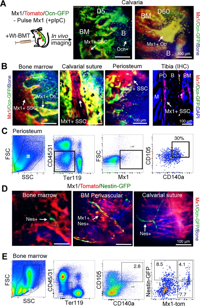
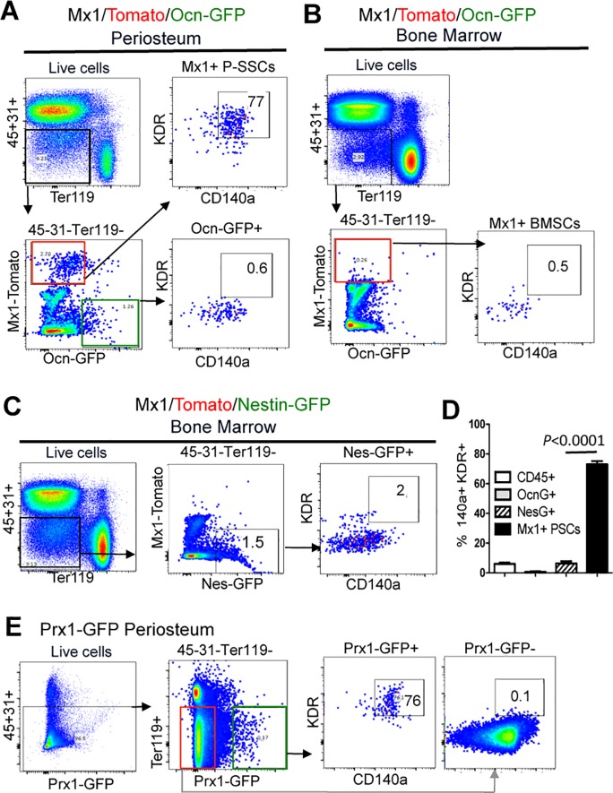
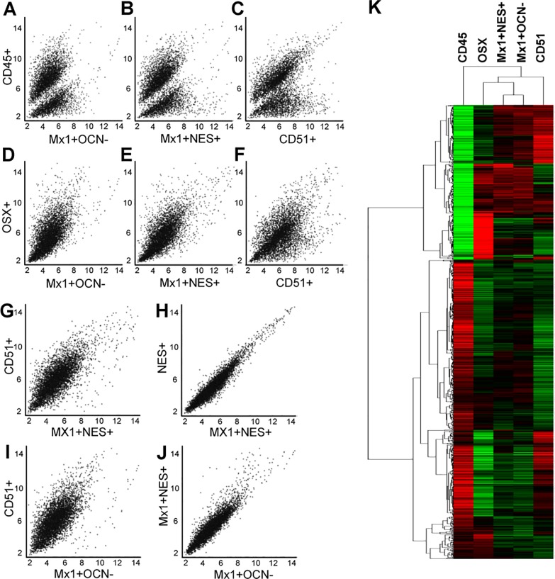
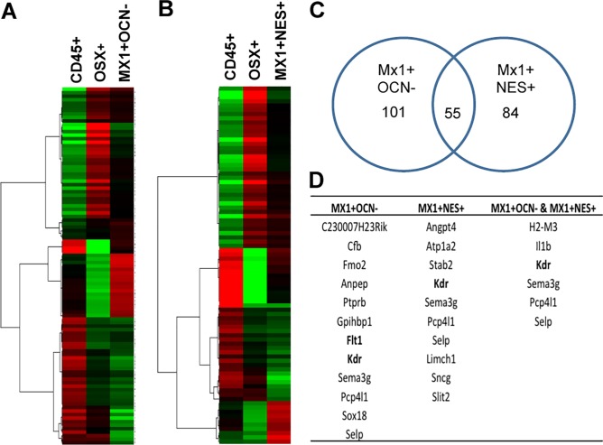
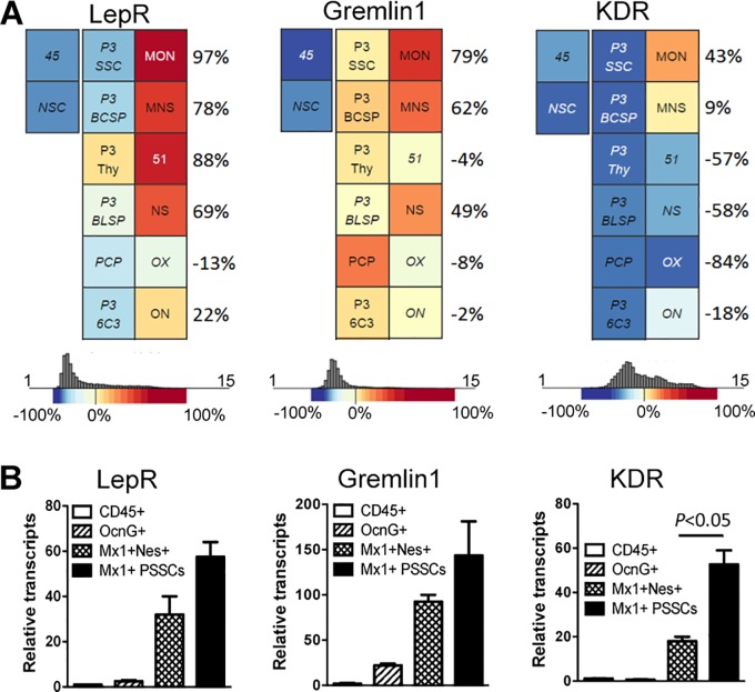
Periosteum and bone marrow (BM) both contain skeletal stem/progenitor cells (SSCs) that participate in fracture repair. However, the functional difference and selective regulatory mechanisms of SSCs in different locations are unknown due to the lack of specific markers. Here, we report a comprehensive gene expression analysis of bone marrow SSCs (BM-SSCs), periosteal SSCs (P-SSCs), and more differentiated osteoprogenitors by using reporter mice expressing Interferon-inducible Mx1 and NestinGFP, previously known SSC markers. We first defined that the BM-SSCs can be enriched by the combination of Mx1 and NestinGFP expression, while endogenous P-SSCs can be isolated by positive selection of Mx1, CD105 and CD140a (known SSC markers) combined with the negative selection of CD45, CD31, and osteocalcinGFP (a mature osteoblast marker). Comparative gene expression analysis with FACS-sorted BM-SSCs, P-SSCs, Osterix+ preosteoblasts, CD51+ stroma cells and CD45+ hematopoietic cells as controls revealed that BM-SSCs and P-SSCs have high similarity with few potential differences without statistical significance. We also found that CD51+ cells are highly heterogeneous and have little overlap with SSCs. This was further supported by the microarray cluster analysis, where the two SSC populations clustered together but are separate from the CD51+ cells. However, when comparing SSC population to controls, we found several genes that are uniquely upregulated in endogenous SSCs. Amongst these genes, we found KDR (aka Flk1 or VEGFR2) to be most interesting and discovered that it is highly and selectively expressed in P-SSCs. This finding suggests that endogenous P-SSCs are functionally very similar to BM-SSCs with undetectable significant differences in gene expression but there are distinct molecular signatures in P-SSCs, which can be useful to specify P-SSC subset in vivo.
骨膜和骨髓(BM)都含有参与骨折修复的骨骼干/祖细胞(SSCs)。然而,由于缺乏特异性标志物,不同位置的SSCs的功能差异和选择性调控机制尚不清楚。在这里,我们通过使用表达干扰素诱导型Mx1和NestinGFP(先前已知的SSC标志物)的报告小鼠,对骨髓SSCs(BM-SSCs)、骨膜SSCs(P-SSCs)以及更分化的骨祖细胞进行了全面的基因表达分析。我们首先确定,BM-SSCs可以通过Mx1和NestinGFP表达的组合来富集,而内源性P-SSCs可以通过Mx1、CD105和CD140a(已知的SSC标志物)的阳性选择与CD45、CD31和骨钙素GFP(一种成熟成骨细胞标志物)的阴性选择相结合来分离。以FACS分选的BM-SSCs、P-SSCs、Osterix+前成骨细胞、CD51+基质细胞和CD45+造血细胞作为对照进行的比较基因表达分析表明,BM-SSCs和P-SSCs具有高度相似性,潜在差异很少且无统计学意义。我们还发现CD51+细胞高度异质性,与SSCs几乎没有重叠。这一点在微阵列聚类分析中得到了进一步支持,其中两个SSC群体聚集在一起,但与CD51+细胞分开。然而,当将SSC群体与对照进行比较时,我们发现了一些在内源性SSCs中独特上调的基因。在这些基因中,我们发现KDR(又名Flk1或VEGFR2)最有趣,并发现它在P-SSCs中高度且选择性地表达。这一发现表明,内源性P-SSCs在功能上与BM-SSCs非常相似,基因表达中未检测到显著差异,但P-SSCs中有独特的分子特征,这可能有助于在体内确定P-SSC亚群。