• 文献检索
  • 文档翻译
  • 深度研究
  • 学术资讯
  • Suppr Zotero 插件Zotero 插件
  • 邀请有礼
  • 套餐&价格
  • 历史记录
应用&插件
Suppr Zotero 插件Zotero 插件浏览器插件Mac 客户端Windows 客户端微信小程序
定价
高级版会员购买积分包购买API积分包
服务
文献检索文档翻译深度研究API 文档MCP 服务
关于我们
关于 Suppr公司介绍联系我们用户协议隐私条款
关注我们

Suppr 超能文献

核心技术专利:CN118964589B侵权必究
粤ICP备2023148730 号-1Suppr @ 2026

文献检索

告别复杂PubMed语法,用中文像聊天一样搜索,搜遍4000万医学文献。AI智能推荐,让科研检索更轻松。

立即免费搜索

文件翻译

保留排版,准确专业,支持PDF/Word/PPT等文件格式,支持 12+语言互译。

免费翻译文档

深度研究

AI帮你快速写综述,25分钟生成高质量综述,智能提取关键信息,辅助科研写作。

立即免费体验

使用正面扫频源光学相干断层扫描血管造影术检测慢性中心性浆液性脉络膜视网膜病变中的静止性I型脉络膜新生血管膜

Detection of Silent Type I Choroidal Neovascular Membrane in Chronic Central Serous Chorioretinopathy Using En Face Swept-Source Optical Coherence Tomography Angiography.

作者信息

Moussa Magdy, Leila Mahmoud, Khalid Hagar, Lolah Mohamed

机构信息

Ophthalmology Department, Faculty of Medicine, Tanta University, Tanta, Egypt.

MEDIC Eye Center, Tanta, Egypt.

出版信息

J Ophthalmol. 2017;2017:6913980. doi: 10.1155/2017/6913980. Epub 2017 Dec 4.

DOI:10.1155/2017/6913980
PMID:29348931
原文链接:https://pmc.ncbi.nlm.nih.gov/articles/PMC5733975/
Abstract

PURPOSE

To evaluate the efficacy of SS-OCTA in the detection of silent CNV secondary to chronic CSCR compared to that of FFA and SS-OCT.

PATIENTS AND METHODS

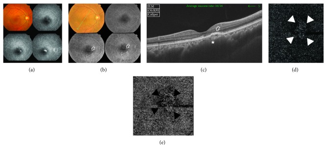
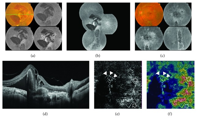
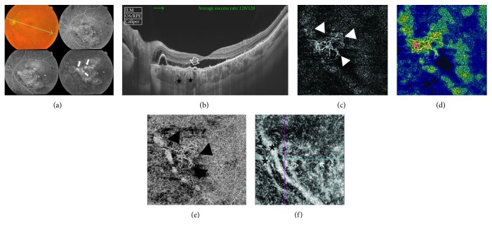
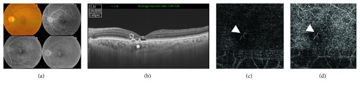
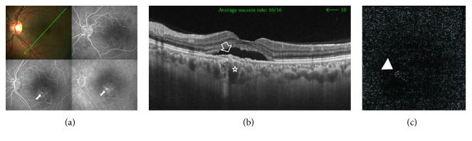
A retrospective observational case series reviewing the clinical data, FFA, SS-OCT, and SS-OCTA images of patients with chronic CSCR, and comparing the findings. SS-OCTA detects the CNV complex and delineates it from the surrounding pathological features of chronic CSCR by utilizing the blood flow detection algorithm, OCTARA, and the ultrahigh-definition B-scan images of the retinal microstructure generated by swept-source technology. The bivariate correlation procedure was used for the calculation of the correlation matrix of the variables tested.

RESULTS

The study included 60 eyes of 40 patients. Mean age was 47.6 years. Mean disease duration was 14.5 months. SS-OCTA detected type 1 CNV in 5 eyes (8.3%). In all 5 eyes, FFA and SS-OCT were inconclusive for CNV. The presence of foveal thinning, opaque material beneath irregular flat PED, and increased choroidal thickness in chronic CSCR constitutes a high-risk profile for progression to CNV development.

CONCLUSION

Silent type 1 CNV is an established complication of chronic CSCR. SS-OCTA is indispensable in excluding CNV especially in high-risk patients and whenever FFA and SS-OCT are inconclusive.

摘要

目的

与荧光素眼底血管造影(FFA)和扫频光学相干断层扫描(SS-OCT)相比,评估扫频光学相干断层扫描血管造影(SS-OCTA)检测慢性中心性浆液性脉络膜视网膜病变(CSCR)继发的隐匿性脉络膜新生血管(CNV)的疗效。

患者与方法

一项回顾性观察病例系列研究,回顾慢性CSCR患者的临床数据、FFA、SS-OCT和SS-OCTA图像,并比较研究结果。SS-OCTA利用血流检测算法OCTARA以及扫频源技术生成的视网膜微观结构的超高分辨率B扫描图像,检测CNV复合体并将其与慢性CSCR的周围病理特征区分开来。采用双变量相关程序计算所测试变量的相关矩阵。

结果

该研究纳入了40例患者的60只眼。平均年龄为47.6岁。平均病程为14.5个月。SS-OCTA在5只眼(8.3%)中检测到1型CNV。在所有5只眼中,FFA和SS-OCT对CNV的诊断均不明确。慢性CSCR中存在黄斑变薄、不规则扁平色素上皮脱离(PED)下方的不透明物质以及脉络膜厚度增加,构成了进展为CNV的高危特征。

结论

隐匿性1型CNV是慢性CSCR的一种既定并发症。SS-OCTA在排除CNV方面不可或缺,尤其是在高危患者以及FFA和SS-OCT诊断不明确时。

https://cdn.ncbi.nlm.nih.gov/pmc/blobs/652b/5733975/975fc781eb49/JOPH2017-6913980.005.jpg
https://cdn.ncbi.nlm.nih.gov/pmc/blobs/652b/5733975/cc32bce45739/JOPH2017-6913980.001.jpg
https://cdn.ncbi.nlm.nih.gov/pmc/blobs/652b/5733975/1ffa18c78c0e/JOPH2017-6913980.002.jpg
https://cdn.ncbi.nlm.nih.gov/pmc/blobs/652b/5733975/0c3070318196/JOPH2017-6913980.003.jpg
https://cdn.ncbi.nlm.nih.gov/pmc/blobs/652b/5733975/314127746043/JOPH2017-6913980.004.jpg
https://cdn.ncbi.nlm.nih.gov/pmc/blobs/652b/5733975/975fc781eb49/JOPH2017-6913980.005.jpg

相似文献

1
Detection of Silent Type I Choroidal Neovascular Membrane in Chronic Central Serous Chorioretinopathy Using En Face Swept-Source Optical Coherence Tomography Angiography.使用正面扫频源光学相干断层扫描血管造影术检测慢性中心性浆液性脉络膜视网膜病变中的静止性I型脉络膜新生血管膜
J Ophthalmol. 2017;2017:6913980. doi: 10.1155/2017/6913980. Epub 2017 Dec 4.
2
Imaging choroidal neovascular membrane using en face swept-source optical coherence tomography angiography.使用正面扫频源光学相干断层扫描血管造影术对脉络膜新生血管膜进行成像。
Clin Ophthalmol. 2017 Oct 13;11:1859-1869. doi: 10.2147/OPTH.S143018. eCollection 2017.
3
Clinical evaluation of neovascular and non-neovascular chronic central serous chorioretinopathy (CSC) diagnosed by swept source optical coherence tomography angiography (SS OCTA).经扫频源光学相干断层扫描血管造影(SS OCTA)诊断的新生血管性和非新生血管性慢性中心性浆液性脉络膜视网膜病变(CSC)的临床评估
Graefes Arch Clin Exp Ophthalmol. 2019 Aug;257(8):1581-1590. doi: 10.1007/s00417-019-04297-z. Epub 2019 Apr 29.
4
En face enhanced-depth swept-source optical coherence tomography features of chronic central serous chorioretinopathy.慢性中心性浆液性脉络膜视网膜病变的光学相干断层扫描特征。
Ophthalmology. 2014 Mar;121(3):719-26. doi: 10.1016/j.ophtha.2013.10.014. Epub 2013 Nov 26.
5
Three-Year Follow-Up Detecting Choroidal Neovascularization with Swept Source Optical Coherence Tomography Angiography (SS-OCTA) after Successful Half-Fluence Photodynamic Therapy for Chronic Central Serous Chorioretinopathy.慢性中心性浆液性脉络膜视网膜病变半量光动力疗法成功后,使用扫频源光学相干断层扫描血管造影(SS-OCTA)进行三年随访以检测脉络膜新生血管形成
Diagnostics (Basel). 2023 Aug 29;13(17):2792. doi: 10.3390/diagnostics13172792.
6
Association of Choroidal Neovascularization and Central Serous Chorioretinopathy With Optical Coherence Tomography Angiography.脉络膜新生血管和中心性浆液性脉络膜视网膜病变与光学相干断层扫描血管造影的关联
JAMA Ophthalmol. 2015 Aug;133(8):899-906. doi: 10.1001/jamaophthalmol.2015.1320.
7
Optical coherence tomography angiography of flat irregular pigment epithelial detachments in central serous chorioretinopathy.中心性浆液性脉络膜视网膜病变中扁平状不规则色素上皮脱离的光相干断层扫描血管造影。
Br J Ophthalmol. 2021 Feb;105(2):233-238. doi: 10.1136/bjophthalmol-2019-315318. Epub 2020 Apr 7.
8
Correlation between Choroidal Neovascularization Shown by OCT Angiography and Choroidal Thickness in Patients with Chronic Central Serous Chorioretinopathy.慢性中心性浆液性脉络膜视网膜病变患者中,光学相干断层扫描血管造影显示的脉络膜新生血管与脉络膜厚度之间的相关性
J Ophthalmol. 2017;2017:3048013. doi: 10.1155/2017/3048013. Epub 2017 Oct 4.
9
Optical coherence tomography angiography findings in fellow eyes of choroidal neovascularisation associated with central serous chorioretinopathy.中心性浆液性脉络膜视网膜病变相关脉络膜新生血管对侧眼的光学相干断层扫描血管造影结果
Br J Ophthalmol. 2021 Sep;105(9):1280-1285. doi: 10.1136/bjophthalmol-2018-313576. Epub 2019 Feb 23.
10
Multimodal Imaging in the Management of Choroidal Neovascularization Secondary to Central Serous Chorioretinopathy.多模态成像在中心性浆液性脉络膜视网膜病变继发脉络膜新生血管管理中的应用
J Clin Med. 2020 Jun 21;9(6):1934. doi: 10.3390/jcm9061934.

引用本文的文献

1
Advances in swept-source optical coherence tomography and optical coherence tomography angiography.扫频源光学相干断层扫描及光学相干断层扫描血管造影术的进展。
Adv Ophthalmol Pract Res. 2022 Nov 25;3(2):67-79. doi: 10.1016/j.aopr.2022.10.005. eCollection 2023 May-Jun.
2
Multimodal Imaging Based Predictors for the Development of Choroidal Neovascularization in Patients with Central Serous Chorioretinopathy.基于多模态成像的中心性浆液性脉络膜视网膜病变患者脉络膜新生血管形成的预测因素
J Clin Med. 2023 Mar 6;12(5):2069. doi: 10.3390/jcm12052069.
3
A customized regimen of intravitreal aflibercept for the treatment of choroidal neovascularization secondary to different chorioretinal diseases.

本文引用的文献

1
Swept-Source Optical Coherence Tomography Angio™ (Topcon Corp, Japan): Technology Review.扫频源光学相干断层扫描血管造影术™(日本拓普康公司):技术综述
Dev Ophthalmol. 2016;56:13-7. doi: 10.1159/000442771. Epub 2016 Mar 15.
2
Vascularization of Irregular Retinal Pigment Epithelial Detachments in Chronic Central Serous Chorioretinopathy Evaluated With OCT Angiography.光学相干断层扫描血管造影评估慢性中心性浆液性脉络膜视网膜病变中不规则视网膜色素上皮脱离的血管化情况
Ophthalmic Surg Lasers Imaging Retina. 2016 Feb;47(2):128-33. doi: 10.3928/23258160-20160126-05.
3
NEW INSIGHT INTO THE MACULAR DEEP VASCULAR PLEXUS IMAGED BY OPTICAL COHERENCE TOMOGRAPHY ANGIOGRAPHY.
用于治疗继发于不同脉络膜视网膜疾病的脉络膜新生血管的定制阿柏西普玻璃体内注射方案。
Int J Retina Vitreous. 2023 Jan 18;9(1):2. doi: 10.1186/s40942-023-00440-5.
4
A Review of Central Serous Chorioretinopathy: Clinical Presentation and Management.中心性浆液性脉络膜视网膜病变综述:临床表现与治疗
Cureus. 2022 Aug 13;14(8):e27965. doi: 10.7759/cureus.27965. eCollection 2022 Aug.
5
Multimodal imaging of macular telangiectasia type 2 in a pediatric patient.一名儿科患者的2型黄斑毛细血管扩张症的多模态成像
Am J Ophthalmol Case Rep. 2021 Apr 1;22:101082. doi: 10.1016/j.ajoc.2021.101082. eCollection 2021 Jun.
6
Central Serous Chorioretinopathy Classification.中心性浆液性脉络膜视网膜病变分类
Pharmaceuticals (Basel). 2020 Dec 30;14(1):26. doi: 10.3390/ph14010026.
7
Assessment of choriocapillary blood flow changes in response to half-dose photodynamic therapy in chronic central serous chorioretinopathy using optical coherence tomography angiography.使用光学相干断层扫描血管造影术评估慢性中心性浆液性脉络膜视网膜病变中脉络膜毛细血管血流对半剂量光动力疗法的反应变化。
BMC Ophthalmol. 2020 Oct 7;20(1):402. doi: 10.1186/s12886-020-01674-9.
8
Optical Coherence Tomography Angiography in Neurodegenerative Diseases: A Review.神经退行性疾病中的光学相干断层扫描血管造影:综述
Eye Brain. 2020 Jul 14;12:73-87. doi: 10.2147/EB.S193026. eCollection 2020.
9
Clinical evaluation of neovascular and non-neovascular chronic central serous chorioretinopathy (CSC) diagnosed by swept source optical coherence tomography angiography (SS OCTA).经扫频源光学相干断层扫描血管造影(SS OCTA)诊断的新生血管性和非新生血管性慢性中心性浆液性脉络膜视网膜病变(CSC)的临床评估
Graefes Arch Clin Exp Ophthalmol. 2019 Aug;257(8):1581-1590. doi: 10.1007/s00417-019-04297-z. Epub 2019 Apr 29.
10
Emerging Applications of Optical Coherence Tomography Angiography (OCTA) in neurological research.光学相干断层扫描血管造影术(OCTA)在神经学研究中的新兴应用。
Eye Vis (Lond). 2018 May 12;5:11. doi: 10.1186/s40662-018-0104-3. eCollection 2018.
光学相干断层扫描血管造影术对黄斑深层血管丛的新见解。
Retina. 2015 Nov;35(11):2347-52. doi: 10.1097/IAE.0000000000000839.
4
Optimization of the split-spectrum amplitude-decorrelation angiography algorithm on a spectral optical coherence tomography system.光谱光学相干断层扫描系统上的分裂谱幅度去相关血管造影算法的优化
Opt Lett. 2015 May 15;40(10):2305-8. doi: 10.1364/OL.40.002305.
5
Loculation of Fluid in the Posterior Choroid in Eyes With Central Serous Chorioretinopathy.中心性浆液性脉络膜视网膜病变患者眼后脉络膜的液体潴留
Am J Ophthalmol. 2015 Dec;160(6):1211-6. doi: 10.1016/j.ajo.2015.08.018. Epub 2015 Aug 20.
6
OCT Angiography Compared to Fluorescein and Indocyanine Green Angiography in Chronic Central Serous Chorioretinopathy.光学相干断层扫描血管造影与荧光素及吲哚菁绿血管造影在慢性中心性浆液性脉络膜视网膜病变中的比较
Invest Ophthalmol Vis Sci. 2015 Aug;56(9):5229-37. doi: 10.1167/iovs.15-17140.
7
Optical coherence tomographic angiography of choroidal neovascularization associated with central serous chorioretinopathy.中心性浆液性脉络膜视网膜病变相关脉络膜新生血管的光学相干断层扫描血管造影
JAMA Ophthalmol. 2015 Oct;133(10):1212-4. doi: 10.1001/jamaophthalmol.2015.2126.
8
Chronic central serous chorioretinopathy imaged by optical coherence tomographic angiography.光学相干断层扫描血管造影成像的慢性中心性浆液性脉络膜视网膜病变。
Am J Ophthalmol. 2015 Sep;160(3):581-587.e1. doi: 10.1016/j.ajo.2015.06.016. Epub 2015 Jun 29.
9
Noninvasive Visualization and Analysis of the Human Parafoveal Capillary Network Using Swept Source OCT Optical Microangiography.使用扫频源光学相干断层扫描光学微血管造影术对人黄斑旁毛细血管网络进行无创可视化和分析。
Invest Ophthalmol Vis Sci. 2015 Jun;56(6):3984-8. doi: 10.1167/iovs.15-16510.
10
Association of Choroidal Neovascularization and Central Serous Chorioretinopathy With Optical Coherence Tomography Angiography.脉络膜新生血管和中心性浆液性脉络膜视网膜病变与光学相干断层扫描血管造影的关联
JAMA Ophthalmol. 2015 Aug;133(8):899-906. doi: 10.1001/jamaophthalmol.2015.1320.