• 文献检索
  • 文档翻译
  • 深度研究
  • 学术资讯
  • Suppr Zotero 插件Zotero 插件
  • 邀请有礼
  • 套餐&价格
  • 历史记录
应用&插件
Suppr Zotero 插件Zotero 插件浏览器插件Mac 客户端Windows 客户端微信小程序
定价
高级版会员购买积分包购买API积分包
服务
文献检索文档翻译深度研究API 文档MCP 服务
关于我们
关于 Suppr公司介绍联系我们用户协议隐私条款
关注我们

Suppr 超能文献

核心技术专利:CN118964589B侵权必究
粤ICP备2023148730 号-1Suppr @ 2026

文献检索

告别复杂PubMed语法,用中文像聊天一样搜索,搜遍4000万医学文献。AI智能推荐,让科研检索更轻松。

立即免费搜索

文件翻译

保留排版,准确专业,支持PDF/Word/PPT等文件格式,支持 12+语言互译。

免费翻译文档

深度研究

AI帮你快速写综述,25分钟生成高质量综述,智能提取关键信息,辅助科研写作。

立即免费体验

在 3T MRI 下活体测量的人类大脑皮质的详细 T1 加权图谱。

Detailed T1-Weighted Profiles from the Human Cortex Measured in Vivo at 3 Tesla MRI.

机构信息

Brain Center Rudolf Magnus, Department of Psychiatry, Brain Division, University Medical Center Utrecht, Utrecht University, HPNR A01.126, Heidelberglaan 100, 3584, CG, Utrecht, The Netherlands.

Radiology Department, Imaging Division, University Medical Center Utrecht, Heidelberglaan 100, 3584, CG, Utrecht, The Netherlands.

出版信息

Neuroinformatics. 2018 Apr;16(2):181-196. doi: 10.1007/s12021-018-9356-2.

DOI:10.1007/s12021-018-9356-2
PMID:29352389
原文链接:https://pmc.ncbi.nlm.nih.gov/articles/PMC5984962/
Abstract

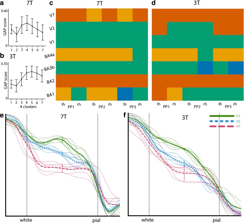
Studies into cortical thickness in psychiatric diseases based on T1-weighted MRI frequently report on aberrations in the cerebral cortex. Due to limitations in image resolution for studies conducted at conventional MRI field strengths (e.g. 3 Tesla (T)) this information cannot be used to establish which of the cortical layers may be implicated. Here we propose a new analysis method that computes one high-resolution average cortical profile per brain region extracting myeloarchitectural information from T1-weighted MRI scans that are routinely acquired at a conventional field strength. To assess this new method, we acquired standard T1-weighted scans at 3 T and compared them with state-of-the-art ultra-high resolution T1-weighted scans optimised for intracortical myelin contrast acquired at 7 T. Average cortical profiles were computed for seven different brain regions. Besides a qualitative comparison between the 3 T scans, 7 T scans, and results from literature, we tested if the results from dynamic time warping-based clustering are similar for the cortical profiles computed from 7 T and 3 T data. In addition, we quantitatively compared cortical profiles computed for V1, V2 and V7 for both 7 T and 3 T data using a priori information on their relative myelin concentration. Although qualitative comparisons show that at an individual level average profiles computed for 7 T have more pronounced features than 3 T profiles the results from the quantitative analyses suggest that average cortical profiles computed from T1-weighted scans acquired at 3 T indeed contain myeloarchitectural information similar to profiles computed from the scans acquired at 7 T. The proposed method therefore provides a step forward to study cortical myeloarchitecture in vivo at conventional magnetic field strength both in health and disease.

摘要

基于 T1 加权 MRI 的精神疾病皮质厚度研究经常报告大脑皮层的异常。由于常规 MRI 场强(例如 3 特斯拉(T))下进行的研究的图像分辨率有限,因此无法使用这些信息来确定哪些皮质层可能受到影响。在这里,我们提出了一种新的分析方法,该方法可以为每个大脑区域计算一个高分辨率的平均皮质轮廓,从常规场强下常规获取的 T1 加权 MRI 扫描中提取髓鞘结构信息。为了评估这种新方法,我们在 3T 处获取了标准 T1 加权扫描,并将其与针对 7T 下皮质内髓鞘对比度优化的最先进超高分辨率 T1 加权扫描进行了比较。为七个不同的大脑区域计算了平均皮质轮廓。除了对 3T 扫描、7T 扫描和文献结果进行定性比较之外,我们还测试了基于动态时间 warping 的聚类的结果是否对于从 7T 和 3T 数据计算出的皮质轮廓相似。此外,我们使用有关其相对髓鞘浓度的先验信息,对来自 7T 和 3T 数据的 V1、V2 和 V7 的皮质轮廓进行了定量比较。尽管定性比较表明,在个体水平上,7T 计算出的平均轮廓比 3T 轮廓具有更明显的特征,但定量分析的结果表明,从 3T 获取的 T1 加权扫描计算出的平均皮质轮廓确实包含与从 7T 获取的扫描计算出的轮廓相似的髓鞘结构信息。因此,该方法为在常规磁场强度下研究健康和疾病中的皮质髓鞘结构提供了一个新的步骤。

https://cdn.ncbi.nlm.nih.gov/pmc/blobs/6bd9/5984962/bb4fe6438fa2/12021_2018_9356_Fig6_HTML.jpg
https://cdn.ncbi.nlm.nih.gov/pmc/blobs/6bd9/5984962/a95985084e2b/12021_2018_9356_Fig1_HTML.jpg
https://cdn.ncbi.nlm.nih.gov/pmc/blobs/6bd9/5984962/d831dc7f594c/12021_2018_9356_Fig2_HTML.jpg
https://cdn.ncbi.nlm.nih.gov/pmc/blobs/6bd9/5984962/3c5d43d70896/12021_2018_9356_Fig3_HTML.jpg
https://cdn.ncbi.nlm.nih.gov/pmc/blobs/6bd9/5984962/0e151c1e4f66/12021_2018_9356_Fig4_HTML.jpg
https://cdn.ncbi.nlm.nih.gov/pmc/blobs/6bd9/5984962/87937517b53d/12021_2018_9356_Fig5_HTML.jpg
https://cdn.ncbi.nlm.nih.gov/pmc/blobs/6bd9/5984962/bb4fe6438fa2/12021_2018_9356_Fig6_HTML.jpg

相似文献

1
Detailed T1-Weighted Profiles from the Human Cortex Measured in Vivo at 3 Tesla MRI.在 3T MRI 下活体测量的人类大脑皮质的详细 T1 加权图谱。
Neuroinformatics. 2018 Apr;16(2):181-196. doi: 10.1007/s12021-018-9356-2.
2
A subject-specific framework for in vivo myeloarchitectonic analysis using high resolution quantitative MRI.使用高分辨率定量 MRI 进行活体骨髓构筑分析的主题特定框架。
Neuroimage. 2016 Jan 15;125:94-107. doi: 10.1016/j.neuroimage.2015.10.001. Epub 2015 Oct 9.
3
Depth-dependent intracortical myelin organization in the living human brain determined by in vivo ultra-high field magnetic resonance imaging.基于活体超高场磁共振成像的人脑皮质内髓鞘组织的深度依赖性研究。
Neuroimage. 2019 Jan 15;185:27-34. doi: 10.1016/j.neuroimage.2018.10.023. Epub 2018 Oct 9.
4
Linking contemporary high resolution magnetic resonance imaging to the von Economo legacy: A study on the comparison of MRI cortical thickness and histological measurements of cortical structure.将当代高分辨率磁共振成像与冯·埃科诺莫的遗产联系起来:一项关于MRI皮质厚度与皮质结构组织学测量比较的研究。
Hum Brain Mapp. 2015 Aug;36(8):3038-46. doi: 10.1002/hbm.22826. Epub 2015 May 19.
5
In vivo characterization of cortical and white matter neuroaxonal pathology in early multiple sclerosis.早期多发性硬化症中皮质和白质神经轴突病变的体内特征分析
Brain. 2017 Nov 1;140(11):2912-2926. doi: 10.1093/brain/awx247.
6
A cytoarchitecture-driven myelin model reveals area-specific signatures in human primary and secondary areas using ultra-high resolution in-vivo brain MRI.基于细胞构筑的髓鞘模型利用超高分辨率体内脑 MRI 揭示人类初级和次级脑区的区域特异性特征。
Neuroimage. 2015 Jul 1;114:71-87. doi: 10.1016/j.neuroimage.2015.04.023. Epub 2015 Apr 18.
7
Effects of MP2RAGE B sensitivity on inter-site T reproducibility and hippocampal morphometry at 7T.7T 下 MP2RAGE B 灵敏度对不同部位 T 值可重复性和海马形态计量学的影响。
Neuroimage. 2021 Jan 1;224:117373. doi: 10.1016/j.neuroimage.2020.117373. Epub 2020 Sep 17.
8
A framework for cortical laminar composition analysis using low-resolution T1 MRI images.利用低分辨率 T1 MRI 图像进行皮质层状结构分析的框架。
Brain Struct Funct. 2019 May;224(4):1457-1467. doi: 10.1007/s00429-019-01848-2. Epub 2019 Feb 19.
9
Analyzing the cortical fine structure as revealed by ex-vivo anatomical MRI.分析离体解剖 MRI 显示的皮质精细结构。
J Comp Neurol. 2023 Dec;531(18):2146-2161. doi: 10.1002/cne.25532. Epub 2023 Jul 31.
10
Intracortical smoothing of small-voxel fMRI data can provide increased detection power without spatial resolution losses compared to conventional large-voxel fMRI data.皮层内平滑小体素 fMRI 数据可以比常规大体素 fMRI 数据提供更高的检测能力,而不会损失空间分辨率。
Neuroimage. 2019 Apr 1;189:601-614. doi: 10.1016/j.neuroimage.2019.01.054. Epub 2019 Jan 25.

引用本文的文献

1
7T MP2RAGE for cortical myelin segmentation: Impact of aging.7T MP2RAGE 用于皮质髓鞘分割:老化的影响。
PLoS One. 2024 Apr 16;19(4):e0299670. doi: 10.1371/journal.pone.0299670. eCollection 2024.
2
Cortical depth profiles in primary visual cortex for illusory and imaginary experiences.初级视皮层中虚幻和想象体验的皮层深度分布。
Nat Commun. 2024 Feb 2;15(1):1002. doi: 10.1038/s41467-024-45065-w.
3
Widespread cortical dyslamination in epilepsy patients with malformations of cortical development.广泛性皮质层发育不良在伴有皮质发育畸形的癫痫患者中。

本文引用的文献

1
Resolution considerations in imaging of the cortical layers.皮质层成像中的分辨率考虑因素。
Neuroimage. 2018 Jan 1;164:112-120. doi: 10.1016/j.neuroimage.2017.02.086. Epub 2017 Mar 6.
2
A multi-modal parcellation of human cerebral cortex.人类大脑皮层的多模态分区
Nature. 2016 Aug 11;536(7615):171-178. doi: 10.1038/nature18933. Epub 2016 Jul 20.
3
Contributions to magnetic susceptibility of brain tissue.对脑组织磁化率的贡献。
Neuroradiology. 2021 Feb;63(2):225-234. doi: 10.1007/s00234-020-02561-2. Epub 2020 Sep 25.
4
Assessing reproducibility in association studies.评估关联研究的可重复性。
Elife. 2019 Apr 25;8:e46757. doi: 10.7554/eLife.46757.
5
Mapping Cortical Laminar Structure in the 3D BigBrain.在 3D BigBrain 中绘制皮质层结构。
Cereb Cortex. 2018 Jul 1;28(7):2551-2562. doi: 10.1093/cercor/bhy074.
NMR Biomed. 2017 Apr;30(4). doi: 10.1002/nbm.3546. Epub 2016 May 30.
4
Lines of Baillarger in vivo and ex vivo: Myelin contrast across lamina at 7T MRI and histology.体内和体外的贝亚尔热线条:7T磁共振成像和组织学下跨层板的髓鞘对比度
Neuroimage. 2016 Jun;133:163-175. doi: 10.1016/j.neuroimage.2016.02.072. Epub 2016 Mar 3.
5
Neurobiological origin of spurious brain morphological changes: A quantitative MRI study.假性脑形态学改变的神经生物学起源:一项定量磁共振成像研究。
Hum Brain Mapp. 2016 May;37(5):1801-15. doi: 10.1002/hbm.23137. Epub 2016 Feb 15.
6
A subject-specific framework for in vivo myeloarchitectonic analysis using high resolution quantitative MRI.使用高分辨率定量 MRI 进行活体骨髓构筑分析的主题特定框架。
Neuroimage. 2016 Jan 15;125:94-107. doi: 10.1016/j.neuroimage.2015.10.001. Epub 2015 Oct 9.
7
Multivariate combination of magnetization transfer, T2* and B0 orientation to study the myelo-architecture of the in vivo human cortex.利用磁化传递、T2*和B0取向的多变量组合研究活体人类皮质的髓鞘结构。
Neuroimage. 2015 Oct 1;119:89-102. doi: 10.1016/j.neuroimage.2015.06.033. Epub 2015 Jun 18.
8
A cytoarchitecture-driven myelin model reveals area-specific signatures in human primary and secondary areas using ultra-high resolution in-vivo brain MRI.基于细胞构筑的髓鞘模型利用超高分辨率体内脑 MRI 揭示人类初级和次级脑区的区域特异性特征。
Neuroimage. 2015 Jul 1;114:71-87. doi: 10.1016/j.neuroimage.2015.04.023. Epub 2015 Apr 18.
9
Cortical thickness gradients in structural hierarchies.结构层次中的皮质厚度梯度。
Neuroimage. 2015 May 1;111:241-50. doi: 10.1016/j.neuroimage.2015.02.036. Epub 2015 Feb 25.
10
High-Resolution Mapping of Myeloarchitecture In Vivo: Localization of Auditory Areas in the Human Brain.体内脊髓结构的高分辨率图谱:人类大脑听觉区域的定位
Cereb Cortex. 2015 Oct;25(10):3394-405. doi: 10.1093/cercor/bhu150. Epub 2014 Jul 3.