Department of Psychiatry, University of Cambridge, Cambridge, UK.
Department of Neurology and Neurosurgery, Montreal Neurological Institute (MNI), McGill University, Montreal, Canada.
Cereb Cortex. 2018 Jul 1;28(7):2551-2562. doi: 10.1093/cercor/bhy074.
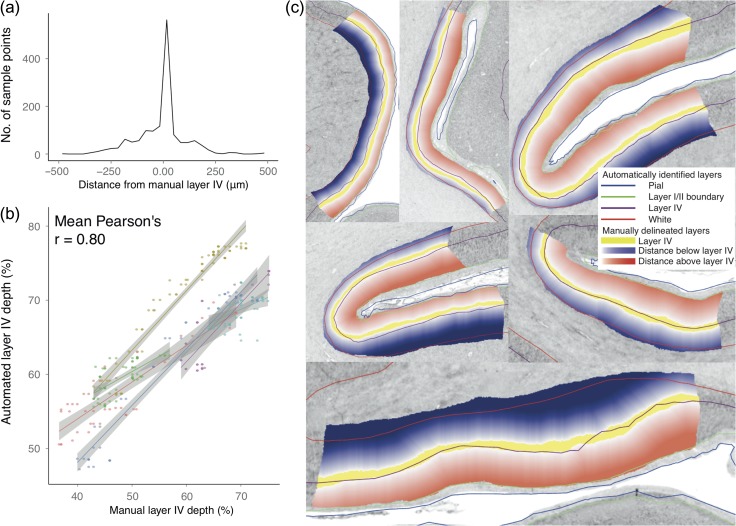
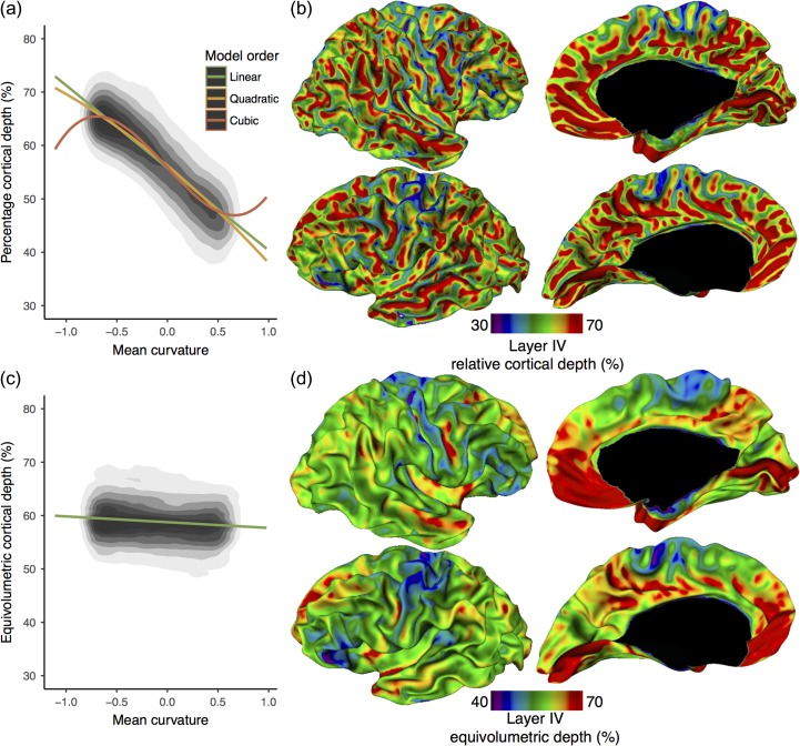
Histological sections offer high spatial resolution to examine laminar architecture of the human cerebral cortex; however, they are restricted by being 2D, hence only regions with sufficiently optimal cutting planes can be analyzed. Conversely, noninvasive neuroimaging approaches are whole brain but have relatively low resolution. Consequently, correct 3D cross-cortical patterns of laminar architecture have never been mapped in histological sections. We developed an automated technique to identify and analyze laminar structure within the high-resolution 3D histological BigBrain. We extracted white matter and pial surfaces, from which we derived histologically verified surfaces at the layer I/II boundary and within layer IV. Layer IV depth was strongly predicted by cortical curvature but varied between areas. This fully automated 3D laminar analysis is an important requirement for bridging high-resolution 2D cytoarchitecture and in vivo 3D neuroimaging. It lays the foundation for in-depth, whole-brain analyses of cortical layering.
组织学切片提供了高空间分辨率,可用于检查人类大脑皮层的层状结构;然而,它们受到 2D 的限制,因此只能分析具有足够最佳切割面的区域。相反,非侵入性的神经影像学方法是整个大脑的,但分辨率相对较低。因此,在组织学切片中从未绘制过正确的 3D 跨皮质层状结构模式。我们开发了一种自动技术,用于识别和分析高分辨率 3D 组织学 BigBrain 中的层状结构。我们提取了白质和软脑膜表面,从中我们得出了在层 I/II 边界和层 IV 内具有组织学验证的表面。层 IV 的深度强烈受到皮质曲率的预测,但在不同区域之间存在差异。这种全自动的 3D 层分析是连接高分辨率 2D 细胞构筑和 3D 体内神经影像学的重要要求。它为皮质分层的深入、全脑分析奠定了基础。