• 文献检索
  • 文档翻译
  • 深度研究
  • 学术资讯
  • Suppr Zotero 插件Zotero 插件
  • 邀请有礼
  • 套餐&价格
  • 历史记录
应用&插件
Suppr Zotero 插件Zotero 插件浏览器插件Mac 客户端Windows 客户端微信小程序
定价
高级版会员购买积分包购买API积分包
服务
文献检索文档翻译深度研究API 文档MCP 服务
关于我们
关于 Suppr公司介绍联系我们用户协议隐私条款
关注我们

Suppr 超能文献

核心技术专利:CN118964589B侵权必究
粤ICP备2023148730 号-1Suppr @ 2026

文献检索

告别复杂PubMed语法,用中文像聊天一样搜索,搜遍4000万医学文献。AI智能推荐,让科研检索更轻松。

立即免费搜索

文件翻译

保留排版,准确专业,支持PDF/Word/PPT等文件格式,支持 12+语言互译。

免费翻译文档

深度研究

AI帮你快速写综述,25分钟生成高质量综述,智能提取关键信息,辅助科研写作。

立即免费体验

屈光不正、血管解剖结构与青光眼性视野缺损之间的相互关系。

The Interrelationship between Refractive Error, Blood Vessel Anatomy, and Glaucomatous Visual Field Loss.

作者信息

Wang Mengyu, Jin Qingying, Wang Hui, Li Dian, Baniasadi Neda, Elze Tobias

机构信息

Schepens Eye Research Institute, Harvard Medical School, Boston, MA, USA.

Jilin University, Changchun, China.

出版信息

Transl Vis Sci Technol. 2018 Jan 18;7(1):4. doi: 10.1167/tvst.7.1.4. eCollection 2018 Jan.

DOI:10.1167/tvst.7.1.4
PMID:29359110
原文链接:https://pmc.ncbi.nlm.nih.gov/articles/PMC5772833/
Abstract

PURPOSE

We quantified the interrelationship between retinal blood vessel (BV) anatomical variation, spherical equivalent (SE) of refractive error, and functional diagnostic parameters in glaucoma to identify optimal parameters for the improvement of optical coherence tomography (OCT) retinal nerve fiber layer thickness (RNFLT) norms.

METHODS

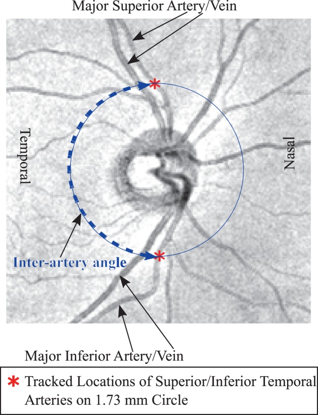
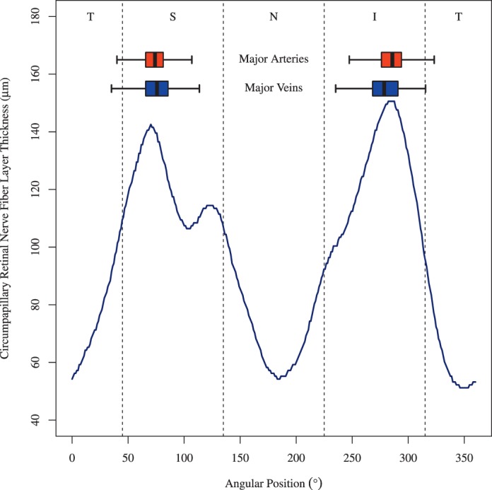
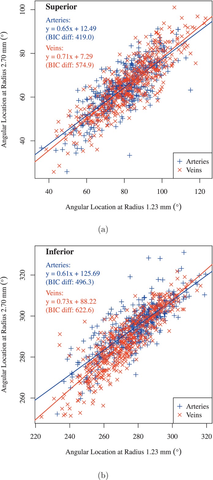
A trained observer marked the intersections of the main superior/inferior temporal arteries and veins with concentric circles around the optic nerve head (ONH) center on fundus images. The interrelationship of BV, SE, and visual field global parameters was analyzed by multivariate regression and model comparison.

RESULTS

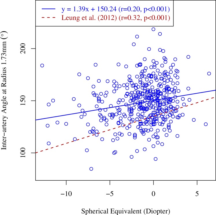
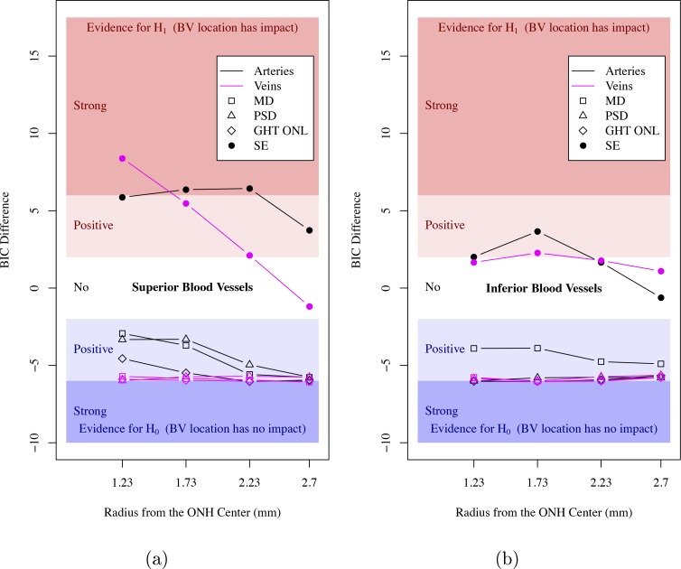
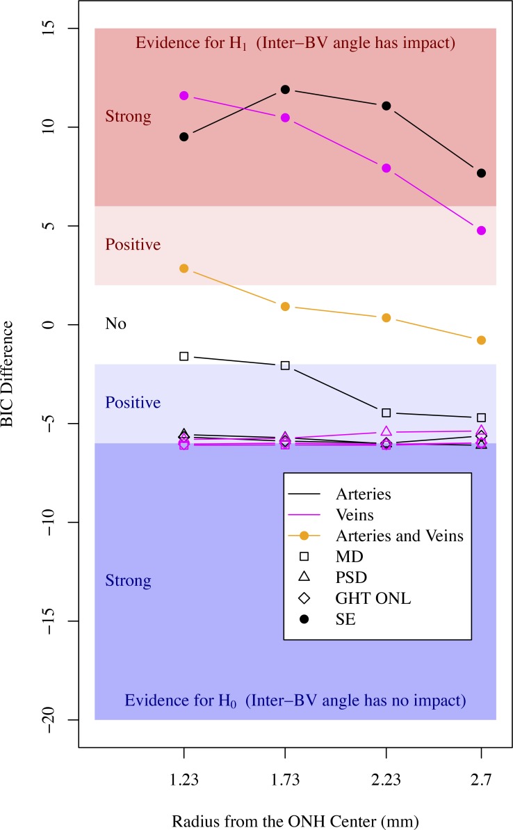
A total of 445 eyes of 445 patients in a large glaucoma practice were selected. Of all investigated BV parameters, interartery angles (IAA) between superior and inferior arteries at a radius of 1.73 mm around the ONH center demonstrated the strongest relationship to SE (Bayesian information criterion difference to null model, 11.9). SE and BV parameters are unrelated to functional parameters, including mean deviation (MD), pattern standard deviation, and glaucoma hemifield test results.

CONCLUSIONS

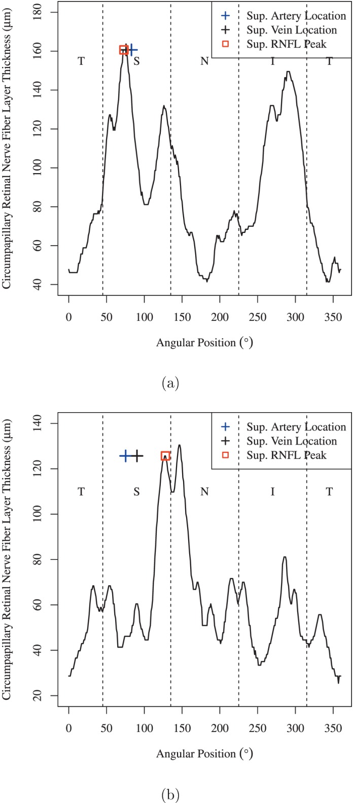
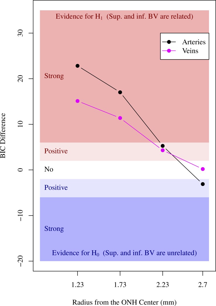
BV locations outside the ONH are sufficiently stable over glaucoma severity to represent individual eye anatomy, and the IAA at 1.73 mm eccentricity is the optimal parameter to be considered for novel OCT RNFLT norms.

TRANSLATIONAL RELEVANCE

Among a large set of BV location parameters, considering IAA may improve RNFLT norms optimally and thereby increase the accuracy of clinical glaucoma diagnosis.

摘要

目的

我们量化了视网膜血管(BV)解剖变异、屈光不正的球镜等效度(SE)与青光眼功能诊断参数之间的相互关系,以确定改善光学相干断层扫描(OCT)视网膜神经纤维层厚度(RNFLT)标准的最佳参数。

方法

一名经过培训的观察者在眼底图像上标记主要颞上/颞下动脉和静脉与围绕视神经乳头(ONH)中心的同心圆的交点。通过多变量回归和模型比较分析BV、SE和视野全局参数之间的相互关系。

结果

在一个大型青光眼诊疗机构中,共选取了445例患者的445只眼睛。在所有研究的BV参数中,围绕ONH中心半径为1.73 mm处的上下动脉之间的动脉间角度(IAA)与SE的关系最为密切(与零模型的贝叶斯信息准则差异为11.9)。SE和BV参数与功能参数无关,包括平均偏差(MD)、模式标准差和青光眼半视野检测结果。

结论

在青光眼严重程度范围内,ONH外部的BV位置足够稳定,可代表个体眼部解剖结构,并且偏心度为1.73 mm处的IAA是新型OCT RNFLT标准应考虑的最佳参数。

转化相关性

在大量BV位置参数中,考虑IAA可能会最佳地改善RNFLT标准,从而提高临床青光眼诊断的准确性。

https://cdn.ncbi.nlm.nih.gov/pmc/blobs/a969/5772833/df081bfe89d7/i2164-2591-7-1-4-f08.jpg
https://cdn.ncbi.nlm.nih.gov/pmc/blobs/a969/5772833/f5624ae2cb98/i2164-2591-7-1-4-f01.jpg
https://cdn.ncbi.nlm.nih.gov/pmc/blobs/a969/5772833/1a1591c29052/i2164-2591-7-1-4-f02.jpg
https://cdn.ncbi.nlm.nih.gov/pmc/blobs/a969/5772833/88d247d888ab/i2164-2591-7-1-4-f03.jpg
https://cdn.ncbi.nlm.nih.gov/pmc/blobs/a969/5772833/a2d92afd5a21/i2164-2591-7-1-4-f04.jpg
https://cdn.ncbi.nlm.nih.gov/pmc/blobs/a969/5772833/50ff8ce10972/i2164-2591-7-1-4-f05.jpg
https://cdn.ncbi.nlm.nih.gov/pmc/blobs/a969/5772833/164aa15cc830/i2164-2591-7-1-4-f06.jpg
https://cdn.ncbi.nlm.nih.gov/pmc/blobs/a969/5772833/15b89bbd1979/i2164-2591-7-1-4-f07.jpg
https://cdn.ncbi.nlm.nih.gov/pmc/blobs/a969/5772833/df081bfe89d7/i2164-2591-7-1-4-f08.jpg

相似文献

1
The Interrelationship between Refractive Error, Blood Vessel Anatomy, and Glaucomatous Visual Field Loss.屈光不正、血管解剖结构与青光眼性视野缺损之间的相互关系。
Transl Vis Sci Technol. 2018 Jan 18;7(1):4. doi: 10.1167/tvst.7.1.4. eCollection 2018 Jan.
2
Ametropia, retinal anatomy, and OCT abnormality patterns in glaucoma. 1. Impacts of refractive error and interartery angle.屈光不正、视网膜解剖和青光眼的 OCT 异常模式。1. 屈光不正和动脉间夹角的影响。
J Biomed Opt. 2017 Dec;22(12):1-11. doi: 10.1117/1.JBO.22.12.121713.
3
Associations between Optic Nerve Head-Related Anatomical Parameters and Refractive Error over the Full Range of Glaucoma Severity.青光眼严重程度全范围内视神经乳头相关解剖学参数与屈光不正之间的关联
Transl Vis Sci Technol. 2017 Jul 18;6(4):9. doi: 10.1167/tvst.6.4.9. eCollection 2017 Jul.
4
Ametropia, retinal anatomy, and OCT abnormality patterns in glaucoma. 2. Impacts of optic nerve head parameters.屈光不正、视网膜解剖和青光眼的 OCT 异常模式。2. 视神经头参数的影响。
J Biomed Opt. 2017 Dec;22(12):1-9. doi: 10.1117/1.JBO.22.12.121714.
5
Quantifying positional variation of retinal blood vessels in glaucoma.定量分析青光眼患者视网膜血管的位置变化。
PLoS One. 2018 Mar 15;13(3):e0193555. doi: 10.1371/journal.pone.0193555. eCollection 2018.
6
Glaucoma diagnostic capabilities of optic nerve head parameters as determined by Cirrus HD optical coherence tomography.Cirrus HD 光学相干断层扫描仪测量的视神经头参数对青光眼的诊断能力。
J Glaucoma. 2012 Sep;21(7):498-504. doi: 10.1097/IJG.0b013e318220dbb7.
7
Retinal nerve fiber layer thickness measured with optical coherence tomography is related to visual function in glaucomatous eyes.通过光学相干断层扫描测量的视网膜神经纤维层厚度与青光眼患者的视觉功能相关。
Ophthalmology. 2003 Nov;110(11):2185-91. doi: 10.1016/S0161-6420(03)00860-1.
8
Application of optical coherence tomography in glaucoma suspect eyes.光学相干断层扫描在青光眼可疑眼中的应用。
Clin Exp Optom. 2012 Jan;95(1):78-88. doi: 10.1111/j.1444-0938.2011.00654.x. Epub 2011 Oct 10.
9
Evaluation of baseline structural factors for predicting glaucomatous visual-field progression using optical coherence tomography, scanning laser polarimetry and confocal scanning laser ophthalmoscopy.采用光学相干断层扫描、扫描激光偏振度测量和共焦激光扫描检眼镜评估预测青光眼视野进展的基线结构因素。
Eye (Lond). 2012 Dec;26(12):1527-35. doi: 10.1038/eye.2012.203. Epub 2012 Oct 12.
10
Optic nerve head and fibre layer imaging for diagnosing glaucoma.用于诊断青光眼的视神经乳头和纤维层成像。
Cochrane Database Syst Rev. 2015 Nov 30;2015(11):CD008803. doi: 10.1002/14651858.CD008803.pub2.

引用本文的文献

1
Retinal Vessel Automatic Segmentation Using SegNet.基于 SegNet 的视网膜血管自动分割。
Comput Math Methods Med. 2022 Mar 26;2022:3117455. doi: 10.1155/2022/3117455. eCollection 2022.
2
Change of Retinal Vessels in Different Sectors of the Parapapillary Area in Primary Open-Angle Glaucoma.原发性开角型青光眼视乳头旁不同区域视网膜血管的变化
Front Med (Lausanne). 2021 Jul 8;8:705829. doi: 10.3389/fmed.2021.705829. eCollection 2021.
3
Towards Automated Eye Diagnosis: An Improved Retinal Vessel Segmentation Framework Using Ensemble Block Matching 3D Filter.

本文引用的文献

1
Associations between Optic Nerve Head-Related Anatomical Parameters and Refractive Error over the Full Range of Glaucoma Severity.青光眼严重程度全范围内视神经乳头相关解剖学参数与屈光不正之间的关联
Transl Vis Sci Technol. 2017 Jul 18;6(4):9. doi: 10.1167/tvst.6.4.9. eCollection 2017 Jul.
2
Relationship Between Central Retinal Vessel Trunk Location and Visual Field Loss in Glaucoma.青光眼患者视网膜中央血管主干位置与视野缺损的关系
Am J Ophthalmol. 2017 Apr;176:53-60. doi: 10.1016/j.ajo.2017.01.001. Epub 2017 Jan 11.
3
Effect of Adjusting Retinal Nerve Fiber Layer Profile to Fovea-Disc Angle Axis on the Thickness and Glaucoma Diagnostic Performance.
迈向自动化眼部诊断:一种使用集成块匹配3D滤波器的改进视网膜血管分割框架。
Diagnostics (Basel). 2021 Jan 12;11(1):114. doi: 10.3390/diagnostics11010114.
4
Inter-Eye Association of Visual Field Defects in Glaucoma and Its Clinical Utility.青光眼视野缺损的双眼关联性及其临床应用。
Transl Vis Sci Technol. 2020 Nov 17;9(12):22. doi: 10.1167/tvst.9.12.22. eCollection 2020 Nov.
5
An Artificial Intelligence Approach to Assess Spatial Patterns of Retinal Nerve Fiber Layer Thickness Maps in Glaucoma.一种用于评估青光眼视网膜神经纤维层厚度图空间模式的人工智能方法。
Transl Vis Sci Technol. 2020 Aug 27;9(9):41. doi: 10.1167/tvst.9.9.41. eCollection 2020 Aug.
6
Sex-Specific Differences in Circumpapillary Retinal Nerve Fiber Layer Thickness.周边视网膜神经纤维层厚度的性别特异性差异。
Ophthalmology. 2020 Mar;127(3):357-368. doi: 10.1016/j.ophtha.2019.09.019. Epub 2019 Sep 25.
7
Compensation of retinal nerve fibre layer thickness as assessed using optical coherence tomography based on anatomical confounders.基于解剖学混杂因素的光学相干断层扫描评估视网膜神经纤维层厚度的补偿。
Br J Ophthalmol. 2020 Feb;104(2):282-290. doi: 10.1136/bjophthalmol-2019-314086. Epub 2019 May 22.
8
Ametropia, retinal anatomy, and OCT abnormality patterns in glaucoma. 1. Impacts of refractive error and interartery angle.屈光不正、视网膜解剖和青光眼的 OCT 异常模式。1. 屈光不正和动脉间夹角的影响。
J Biomed Opt. 2017 Dec;22(12):1-11. doi: 10.1117/1.JBO.22.12.121713.
调整视网膜神经纤维层轮廓至中央凹-视盘角轴对厚度及青光眼诊断性能的影响
Am J Ophthalmol. 2016 Jan;161:12-21.e1-2. doi: 10.1016/j.ajo.2015.09.019. Epub 2015 Sep 24.
4
Influence of the disc-fovea angle on limits of RNFL variability and glaucoma discrimination.盘-黄斑角度对视网膜神经纤维层厚度变异性及青光眼鉴别诊断界值的影响
Invest Ophthalmol Vis Sci. 2014 Oct 9;55(11):7332-42. doi: 10.1167/iovs.14-14962.
5
Improvement of diagnostic performance regarding retinal nerve fiber layer defect using shifting of the normative database according to vessel position.根据血管位置调整规范数据库以提高视网膜神经纤维层缺损的诊断性能。
Invest Ophthalmol Vis Sci. 2014 Jul 29;55(8):5116-24. doi: 10.1167/iovs.14-14630.
6
The foveal position relative to the optic disc and the retinal nerve fiber layer thickness profile in myopia.近视患者黄斑中心凹与视盘的相对位置及视网膜神经纤维层厚度分布。
Invest Ophthalmol Vis Sci. 2014 Mar 10;55(3):1419-26. doi: 10.1167/iovs.13-13604.
7
Correlation between retinal vessel density profile and circumpapillary RNFL thickness measured with Fourier-domain optical coherence tomography.频域光相干断层扫描测量视网膜血管密度与视盘周围神经纤维层厚度的相关性。
Br J Ophthalmol. 2014 Apr;98(4):538-43. doi: 10.1136/bjophthalmol-2013-303910. Epub 2014 Jan 3.
8
Retinal blood vessel positional shifts and glaucoma progression.视网膜血管位置偏移与青光眼进展。
Ophthalmology. 2014 Apr;121(4):842-8. doi: 10.1016/j.ophtha.2013.11.002. Epub 2013 Dec 15.
9
Relationship between position of peak retinal nerve fiber layer thickness and retinal arteries on sectoral retinal nerve fiber layer thickness.节段性视网膜神经纤维层厚度与视网膜动脉在视网膜神经纤维层最厚处位置的关系。
Invest Ophthalmol Vis Sci. 2013 Aug 13;54(8):5481-8. doi: 10.1167/iovs.12-11008.
10
Retinal nerve fiber layer imaging with spectral-domain optical coherence tomography: interpreting the RNFL maps in healthy myopic eyes.频域光学相干断层扫描的视网膜神经纤维层成像:解读健康近视眼中的 RNFL 图。
Invest Ophthalmol Vis Sci. 2012 Oct 17;53(11):7194-200. doi: 10.1167/iovs.12-9726.