• 文献检索
  • 文档翻译
  • 深度研究
  • 学术资讯
  • Suppr Zotero 插件Zotero 插件
  • 邀请有礼
  • 套餐&价格
  • 历史记录
应用&插件
Suppr Zotero 插件Zotero 插件浏览器插件Mac 客户端Windows 客户端微信小程序
定价
高级版会员购买积分包购买API积分包
服务
文献检索文档翻译深度研究API 文档MCP 服务
关于我们
关于 Suppr公司介绍联系我们用户协议隐私条款
关注我们

Suppr 超能文献

核心技术专利:CN118964589B侵权必究
粤ICP备2023148730 号-1Suppr @ 2026

文献检索

告别复杂PubMed语法,用中文像聊天一样搜索,搜遍4000万医学文献。AI智能推荐,让科研检索更轻松。

立即免费搜索

文件翻译

保留排版,准确专业,支持PDF/Word/PPT等文件格式,支持 12+语言互译。

免费翻译文档

深度研究

AI帮你快速写综述,25分钟生成高质量综述,智能提取关键信息,辅助科研写作。

立即免费体验

原发性开角型青光眼视乳头旁不同区域视网膜血管的变化

Change of Retinal Vessels in Different Sectors of the Parapapillary Area in Primary Open-Angle Glaucoma.

作者信息

Cheng Jingyi, Zhao Hongmei, Jiang Chunhui, Kong Xiangmei, Sun Xinghuai

机构信息

Department of Ophthalmology and Visual Science, Eye, Ear, Nose and Throat Hospital, Fudan University, Shanghai, China.

Key Laboratory of Myopia, Ministry of Health, Fudan University, Shanghai, China.

出版信息

Front Med (Lausanne). 2021 Jul 8;8:705829. doi: 10.3389/fmed.2021.705829. eCollection 2021.

DOI:10.3389/fmed.2021.705829
PMID:34307429
原文链接:https://pmc.ncbi.nlm.nih.gov/articles/PMC8295556/
Abstract

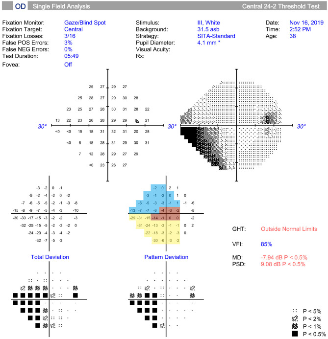
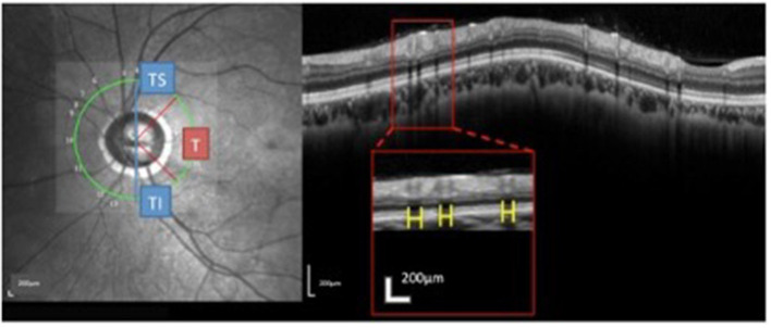
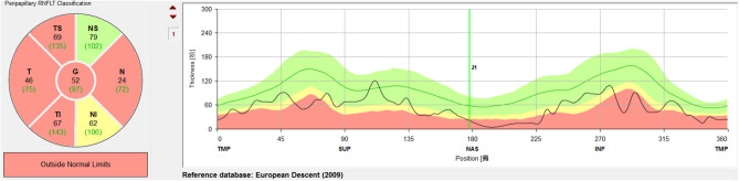
To investigate the changes in the retinal vessels (RVs) in different sectors in patients with primary open-angle glaucoma (POAG), and their possible correlations with retinal nerve fiber layer thickness (RNFLT) and visual-field defects in the temporal parapapillary region. The RV diameters, RNFLTs, and visual-field parameters were measured. The temporal parapapillary region was divided into the temporal (T, 315°-45°), temporal superior (TS, 45°-90°), and temporal inferior sectors (TI, 270°-315°). The changes in the RV diameters in each sector were determined, and their relationships with RNFLT, the mean deviation (MD), and visual field sensitivity (VFS) were examined. Fifty POAG patients (50 eyes) and 50 healthy subjects (50 eyes) were included. Compared with the healthy subjects, the POAG group had a significantly smaller accumulated parapapillary RV diameter ( < 0.001), which was positively correlated with the MD and RNFLT. When the different temporal sectors were examined, the accumulated RV diameters were significantly smaller in the POAG group than in the healthy controls in the TI and T sectors, but not in the TS sector. The accumulated diameters in the TI and T sectors were correlated with the corresponding RNFLTs (all < 0.05), but only the accumulated diameter in the TI sector was correlated with the VFS. In POAG, the changes in the RVs differed between different temporal sectors, with the most prominent changes occurring in the TI and T sectors.

摘要

为研究原发性开角型青光眼(POAG)患者不同象限视网膜血管(RVs)的变化,及其与颞侧视乳头旁区域视网膜神经纤维层厚度(RNFLT)和视野缺损的可能相关性。测量了RV直径、RNFLT和视野参数。将颞侧视乳头旁区域分为颞侧(T,315° - 45°)、颞上(TS,45° - 90°)和颞下象限(TI,270° - 315°)。确定每个象限RV直径的变化,并检查其与RNFLT、平均偏差(MD)和视野敏感度(VFS)的关系。纳入50例POAG患者(50只眼)和50名健康受试者(50只眼)。与健康受试者相比,POAG组视乳头旁RV累积直径显著更小(<0.001),且与MD和RNFLT呈正相关。检查不同颞侧象限时,POAG组TI和T象限的RV累积直径显著小于健康对照组,而TS象限则不然。TI和T象限的累积直径与相应的RNFLT相关(均<0.05),但仅TI象限的累积直径与VFS相关。在POAG中,不同颞侧象限的RV变化不同,TI和T象限变化最为显著。

https://cdn.ncbi.nlm.nih.gov/pmc/blobs/38b1/8295556/461b38f8d5e1/fmed-08-705829-g0003.jpg
https://cdn.ncbi.nlm.nih.gov/pmc/blobs/38b1/8295556/41fe259a1265/fmed-08-705829-g0001.jpg
https://cdn.ncbi.nlm.nih.gov/pmc/blobs/38b1/8295556/d20a29971946/fmed-08-705829-g0002.jpg
https://cdn.ncbi.nlm.nih.gov/pmc/blobs/38b1/8295556/461b38f8d5e1/fmed-08-705829-g0003.jpg
https://cdn.ncbi.nlm.nih.gov/pmc/blobs/38b1/8295556/41fe259a1265/fmed-08-705829-g0001.jpg
https://cdn.ncbi.nlm.nih.gov/pmc/blobs/38b1/8295556/d20a29971946/fmed-08-705829-g0002.jpg
https://cdn.ncbi.nlm.nih.gov/pmc/blobs/38b1/8295556/461b38f8d5e1/fmed-08-705829-g0003.jpg

相似文献

1
Change of Retinal Vessels in Different Sectors of the Parapapillary Area in Primary Open-Angle Glaucoma.原发性开角型青光眼视乳头旁不同区域视网膜血管的变化
Front Med (Lausanne). 2021 Jul 8;8:705829. doi: 10.3389/fmed.2021.705829. eCollection 2021.
2
[The function-structure impairment pattern of optic nerves in primary open-angle glaucoma and normal-tension glaucoma].[原发性开角型青光眼和正常眼压性青光眼视神经的功能-结构损害模式]
Zhonghua Yan Ke Za Zhi. 2018 Nov 11;54(11):811-819. doi: 10.3760/cma.j.issn.0412-4081.2018.11.004.
3
Glaucoma Diagnostic Capability of Circumpapillary Retinal Nerve Fiber Layer Thickness in Circle Scans With Different Diameters.不同直径环形扫描中环乳头视网膜神经纤维层厚度的青光眼诊断能力
J Glaucoma. 2017 Apr;26(4):335-342. doi: 10.1097/IJG.0000000000000610.
4
Parapapillary retinal vessel diameter in normal and glaucoma eyes. II. Correlations.正常眼和青光眼眼中视乳头旁视网膜血管直径。II. 相关性
Invest Ophthalmol Vis Sci. 1989 Jul;30(7):1604-11.
5
[The retinal nerve fiber layer in normal and glaucomatous eyes. II. Correlations].[正常眼和青光眼眼中的视网膜神经纤维层。II. 相关性]
Klin Monbl Augenheilkd. 1989 Nov;195(5):308-14. doi: 10.1055/s-2008-1050044.
6
Deep Retinal Layer Microvasculature Dropout Detected by the Optical Coherence Tomography Angiography in Glaucoma.光学相干断层扫描血管造影术检测青光眼患者视网膜深层微血管缺失
Ophthalmology. 2016 Dec;123(12):2509-2518. doi: 10.1016/j.ophtha.2016.09.002. Epub 2016 Oct 18.
7
The Relationship Between Peripapillary Vascular Density and Visual Field Sensitivity in Primary Open-Angle and Angle-Closure Glaucoma.原发性开角型青光眼和闭角型青光眼的视盘周围血管密度与视野敏感性的关系。
Invest Ophthalmol Vis Sci. 2018 Dec 3;59(15):5862-5867. doi: 10.1167/iovs.18-25423.
8
Parapapillary retinal vessel diameter in normal and glaucoma eyes. I. Morphometric data.正常眼和青光眼眼中视乳头旁视网膜血管直径。I. 形态测量数据。
Invest Ophthalmol Vis Sci. 1989 Jul;30(7):1599-603.
9
[Analysis of morphologic changes of lamina cribrosa in primary open angle glaucoma using enhanced depth imaging optical coherence tomography].[使用增强深度成像光学相干断层扫描分析原发性开角型青光眼筛板的形态学变化]
Zhonghua Yan Ke Za Zhi. 2016 Jun 11;52(6):422-8. doi: 10.3760/cma.j.issn.0412-4081.2016.06.006.
10
Pattern of peripapillary capillary density loss in ischemic optic neuropathy compared to that in primary open-angle glaucoma.与原发性开角型青光眼相比,缺血性视神经病变中视乳头周围毛细血管密度丧失的模式。
PLoS One. 2018 Jan 10;13(1):e0189237. doi: 10.1371/journal.pone.0189237. eCollection 2018.

引用本文的文献

1
Retinal Vessel Diameter Reductions Are Associated with Retinal Ganglion Cell Dysfunction, Thinning of the Ganglion Cell and Inner Plexiform Layers, and Decreased Visual Field Global Indices in Glaucoma Suspects.视网膜血管直径减小与青光眼可疑患者的视网膜神经节细胞功能障碍、神经节细胞层和内网状层变薄以及视野全局指标降低有关。
Diagnostics (Basel). 2025 Jul 3;15(13):1700. doi: 10.3390/diagnostics15131700.
2
Is Primary Open-Angle Glaucoma a Vascular Disease? Assessment of the Relationship between Retinal Arteriolar Morphology and Glaucoma Severity Using Adaptive Optics.原发性开角型青光眼是一种血管疾病吗?利用自适应光学评估视网膜小动脉形态与青光眼严重程度之间的关系。
J Clin Med. 2024 Jan 15;13(2):478. doi: 10.3390/jcm13020478.

本文引用的文献

1
The Interrelationship between Refractive Error, Blood Vessel Anatomy, and Glaucomatous Visual Field Loss.屈光不正、血管解剖结构与青光眼性视野缺损之间的相互关系。
Transl Vis Sci Technol. 2018 Jan 18;7(1):4. doi: 10.1167/tvst.7.1.4. eCollection 2018 Jan.
2
Ametropia, retinal anatomy, and OCT abnormality patterns in glaucoma. 1. Impacts of refractive error and interartery angle.屈光不正、视网膜解剖和青光眼的 OCT 异常模式。1. 屈光不正和动脉间夹角的影响。
J Biomed Opt. 2017 Dec;22(12):1-11. doi: 10.1117/1.JBO.22.12.121713.
3
Structural and Functional Evaluations for the Early Detection of Glaucoma.
青光眼早期检测的结构和功能评估
Expert Rev Ophthalmol. 2016;11(5):367-376. doi: 10.1080/17469899.2016.1229599. Epub 2016 Sep 14.
4
Retinal vessel diameter measurements by spectral domain optical coherence tomography.通过光谱域光学相干断层扫描测量视网膜血管直径
Graefes Arch Clin Exp Ophthalmol. 2015 Apr;253(4):499-509. doi: 10.1007/s00417-014-2715-2. Epub 2014 Aug 17.
5
Retinal blood vessel positional shifts and glaucoma progression.视网膜血管位置偏移与青光眼进展。
Ophthalmology. 2014 Apr;121(4):842-8. doi: 10.1016/j.ophtha.2013.11.002. Epub 2013 Dec 15.
6
Regional variation in human retinal vessel oxygen saturation.人体视网膜血管血氧饱和度的区域性差异。
Exp Eye Res. 2013 Aug;113:143-7. doi: 10.1016/j.exer.2013.06.001. Epub 2013 Jun 18.
7
Correlation of optic disc morphology and ocular perfusion parameters in patients with primary open angle glaucoma.原发性开角型青光眼患者视盘形态与眼血流灌注参数的相关性。
Acta Ophthalmol. 2011 Nov;89(7):e544-9. doi: 10.1111/j.1755-3768.2011.02175.x. Epub 2011 May 23.
8
Relationship of retinal vascular caliber with retinal nerve fiber layer thickness: the singapore malay eye study.视网膜血管管径与视网膜神经纤维层厚度的关系:新加坡马来人眼研究
Invest Ophthalmol Vis Sci. 2009 Sep;50(9):4091-6. doi: 10.1167/iovs.09-3444. Epub 2009 May 14.
9
Regional relationship between retinal nerve fiber layer thickness and corresponding visual field sensitivity in glaucomatous eyes.青光眼患者视网膜神经纤维层厚度与相应视野敏感度之间的区域关系。
Arch Ophthalmol. 2008 Nov;126(11):1500-6. doi: 10.1001/archopht.126.11.1500.
10
Evidence of retinal vascular narrowing in glaucomatous eyes in an Asian population.亚洲人群青光眼患者视网膜血管狭窄的证据。
Invest Ophthalmol Vis Sci. 2008 Dec;49(12):5397-402. doi: 10.1167/iovs.08-2142. Epub 2008 Aug 21.