• 文献检索
  • 文档翻译
  • 深度研究
  • 学术资讯
  • Suppr Zotero 插件Zotero 插件
  • 邀请有礼
  • 套餐&价格
  • 历史记录
应用&插件
Suppr Zotero 插件Zotero 插件浏览器插件Mac 客户端Windows 客户端微信小程序
定价
高级版会员购买积分包购买API积分包
服务
文献检索文档翻译深度研究API 文档MCP 服务
关于我们
关于 Suppr公司介绍联系我们用户协议隐私条款
关注我们

Suppr 超能文献

核心技术专利:CN118964589B侵权必究
粤ICP备2023148730 号-1Suppr @ 2026

文献检索

告别复杂PubMed语法,用中文像聊天一样搜索,搜遍4000万医学文献。AI智能推荐,让科研检索更轻松。

立即免费搜索

文件翻译

保留排版,准确专业,支持PDF/Word/PPT等文件格式,支持 12+语言互译。

免费翻译文档

深度研究

AI帮你快速写综述,25分钟生成高质量综述,智能提取关键信息,辅助科研写作。

立即免费体验

视网膜神经纤维层厚度的最佳双眼差异阈值可预测多发性硬化症中的单侧视神经病变。

Optimal Intereye Difference Thresholds in Retinal Nerve Fiber Layer Thickness for Predicting a Unilateral Optic Nerve Lesion in Multiple Sclerosis.

机构信息

Departments of Neurology (RCN, SLG, and LJB), Population Health (LJB), and Ophthalmology (SLG and LJB), New York University School of Medicine, New York, New York; Department of Neurology (TCF and EMF), Dell Medical School at University of Texas Austin, Austin, Texas; Department of Neurology (PAC), Johns Hopkins University School of Medicine, Baltimore, Maryland; Clinical Development Department (CCV), Biogen, Cambridge, Massachusetts; and Clinical Development Group (DC), Fulcrum Therapeutics, Cambridge, Massachusetts.

出版信息

J Neuroophthalmol. 2018 Dec;38(4):451-458. doi: 10.1097/WNO.0000000000000629.

DOI:10.1097/WNO.0000000000000629
PMID:29384802
原文链接:https://pmc.ncbi.nlm.nih.gov/articles/PMC8845082/
Abstract

BACKGROUND

The optic nerve is a frequent site for involvement in multiple sclerosis (MS). Optical coherence tomography (OCT) detects thinning of the retinal nerve fiber layer (RNFL) in eyes of patients with MS and in those meeting criteria for clinically or radiologically isolated demyelinating syndromes. Current international diagnostic criteria for MS do not include the optic nerve as an imaging lesion site despite the high prevalence of acute optic neuritis (ON), or occult optic neuropathy, among early MS and clinically isolated syndrome patients; as well as most MS patients over the course of the disease. We sought to determine optimal thresholds for intereye difference in peripapillary RNFL thickness that are most predictive of a unilateral optic nerve lesion.

METHODS

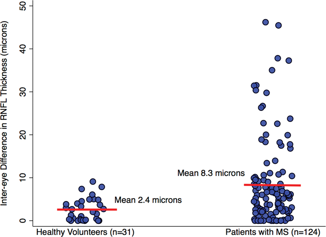
We analyzed spectral domain OCT data of 31 healthy volunteers and 124 patients with MS at a single center as part of an ongoing collaborative investigation of visual outcomes. Intereye differences in peripapillary (360°) RNFL thickness were calculated as the absolute value of the difference. First, we determined the 95th percentile value of intereye difference for the healthy volunteers. This value was applied to the convenience sample group of MS patients as a validation cohort determining how well this threshold could distinguish patients with vs without a history of unilateral ON. The relation of intereye differences in peripapillary RNFL thickness to binocular low-contrast letter acuity scores was also examined.

RESULTS

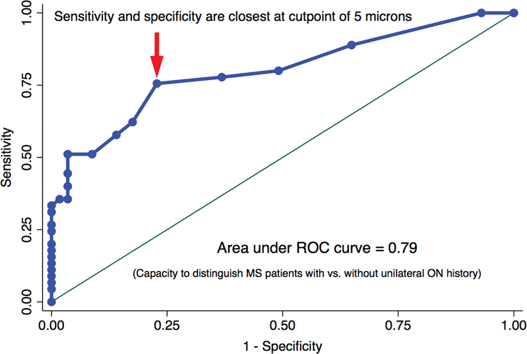
Among healthy volunteer participants (n = 31), the 95th percentile value for intereye difference (upper boundary of expected for normal controls) was 6.0 μm. This value was applied to the convenience sample group of MS patients (n = 124, validation cohort). Positive predictive value, negative predictive value, sensitivity, and specificity for identifying MS patients with a history of unilateral ON were calculated for the 6-μm threshold value in a 2 × 2 table analysis with the application of χ tests (P < 0.0001). The 6-μm threshold was predictive of worse binocular low-contrast acuity scores at 2.5% (P = 0.03) and 1.25% (P = 0.002 by linear regression analyses). A receiver operating characteristic curve analysis demonstrated an optimal intereye difference threshold of 5 μm for identifying unilateral ON in the MS cohort.

CONCLUSIONS

An intereye difference of 5-6 μm in RNFL thickness is a robust structural threshold for identifying the presence of a unilateral optic nerve lesion in MS.

摘要

背景

视神经是多发性硬化症(MS)常累及的部位。光学相干断层扫描(OCT)可检测到 MS 患者和符合临床或放射学孤立脱髓鞘综合征标准的患者的视网膜神经纤维层(RNFL)变薄。尽管急性视神经炎(ON)或隐匿性视神经病变在早期 MS 和临床孤立综合征患者以及大多数 MS 患者中均较为常见,但目前 MS 的国际诊断标准并未将视神经作为影像学病变部位。我们旨在确定最能预测单侧视神经病变的视盘周围 RNFL 厚度的眼间差异的最佳阈值。

方法

我们分析了单中心的 31 名健康志愿者和 124 名 MS 患者的光谱域 OCT 数据,这是一项正在进行的视觉结果合作研究的一部分。视盘周围(360°)RNFL 厚度的眼间差异通过差异的绝对值计算。首先,我们确定了健康志愿者眼间差异的第 95 百分位值。该值应用于 MS 患者的便利样本组作为验证队列,以确定该阈值能在多大程度上区分有单侧 ON 病史和无单侧 ON 病史的患者。我们还检查了视盘周围 RNFL 厚度的眼间差异与双眼低对比度字母视力评分之间的关系。

结果

在健康志愿者参与者(n = 31)中,眼间差异的第 95 百分位值(正常对照组的预期上限)为 6.0 μm。该值应用于 MS 患者的便利样本组(n = 124,验证队列)。通过 χ 检验(P < 0.0001)对 2 × 2 表分析中的 6 μm 阈值值进行了阳性预测值、阴性预测值、敏感性和特异性的计算,以识别有单侧 ON 病史的 MS 患者。线性回归分析表明,6 μm 阈值与双眼低对比度视力评分更差相关(2.5%:P = 0.03;1.25%:P = 0.002)。受试者工作特征曲线分析表明,MS 队列中识别单侧 ON 的最佳眼间差异阈值为 5 μm。

结论

RNFL 厚度的眼间差异为 5-6 μm 是识别 MS 中单侧视神经病变的可靠结构阈值。

https://cdn.ncbi.nlm.nih.gov/pmc/blobs/db35/8845082/94ab55f9f14d/nihms-1764547-f0002.jpg
https://cdn.ncbi.nlm.nih.gov/pmc/blobs/db35/8845082/128458b1e6c2/nihms-1764547-f0001.jpg
https://cdn.ncbi.nlm.nih.gov/pmc/blobs/db35/8845082/94ab55f9f14d/nihms-1764547-f0002.jpg
https://cdn.ncbi.nlm.nih.gov/pmc/blobs/db35/8845082/128458b1e6c2/nihms-1764547-f0001.jpg
https://cdn.ncbi.nlm.nih.gov/pmc/blobs/db35/8845082/94ab55f9f14d/nihms-1764547-f0002.jpg

相似文献

1
Optimal Intereye Difference Thresholds in Retinal Nerve Fiber Layer Thickness for Predicting a Unilateral Optic Nerve Lesion in Multiple Sclerosis.视网膜神经纤维层厚度的最佳双眼差异阈值可预测多发性硬化症中的单侧视神经病变。
J Neuroophthalmol. 2018 Dec;38(4):451-458. doi: 10.1097/WNO.0000000000000629.
2
Optimal intereye difference thresholds by optical coherence tomography in multiple sclerosis: An international study.光学相干断层扫描在多发性硬化症中的最佳眼间差异阈值:一项国际研究。
Ann Neurol. 2019 May;85(5):618-629. doi: 10.1002/ana.25462. Epub 2019 Apr 10.
3
Relation of visual function to retinal nerve fiber layer thickness in multiple sclerosis.多发性硬化症中视觉功能与视网膜神经纤维层厚度的关系
Ophthalmology. 2006 Feb;113(2):324-32. doi: 10.1016/j.ophtha.2005.10.040. Epub 2006 Jan 10.
4
Retinal imaging by laser polarimetry and optical coherence tomography evidence of axonal degeneration in multiple sclerosis.通过激光偏振imetry和光学相干断层扫描进行视网膜成像:多发性硬化症中轴突退变的证据
Arch Neurol. 2008 Jul;65(7):924-8. doi: 10.1001/archneur.65.7.924.
5
Optimization of spectral domain optical coherence tomography and visual evoked potentials to identify unilateral optic neuritis.优化光谱域光学相干断层扫描和视觉诱发电位以识别单侧视神经炎。
Mult Scler Relat Disord. 2020 Jun;41:101988. doi: 10.1016/j.msard.2020.101988. Epub 2020 Feb 7.
6
Changes in oxygen saturation and the retinal nerve fibre layer in patients with optic neuritis associated with multiple sclerosis in a 6-month follow-up.视神经炎相关多发性硬化患者在 6 个月随访中氧饱和度和视网膜神经纤维层的变化。
Acta Ophthalmol. 2020 Dec;98(8):841-847. doi: 10.1111/aos.14463. Epub 2020 May 12.
7
[Clinical investigation of optic coherence tomography in evaluating the impairment of optic nerve secondary to multiple sclerosis].光学相干断层扫描在评估继发于多发性硬化症的视神经损伤中的临床研究
Zhonghua Yan Ke Za Zhi. 2014 Dec;50(12):900-5.
8
Retinal and Optic Nerve Degeneration in Patients with Multiple Sclerosis Followed up for 5 Years.多发性硬化症患者随访 5 年的视网膜和视神经变性。
Ophthalmology. 2017 May;124(5):688-696. doi: 10.1016/j.ophtha.2017.01.005. Epub 2017 Feb 7.
9
Retinal nerve fiber layer and ganglion cell layer changes on optical coherence tomography in early multiple sclerosis and optic neuritis cases.早期多发性硬化症和视神经炎病例中光学相干断层扫描显示的视网膜神经纤维层和神经节细胞层变化
Indian J Ophthalmol. 2018 Jan;66(1):114-119. doi: 10.4103/ijo.IJO_539_17.
10
[Retinal atrophy using optical coherence tomography (OCT) in 15 patients with multiple sclerosis and comparison with healthy subjects].[利用光学相干断层扫描(OCT)对15例多发性硬化症患者视网膜萎缩情况的研究及与健康受试者的比较]
Rev Neurol (Paris). 2008 Nov;164(11):927-34. doi: 10.1016/j.neurol.2008.03.008. Epub 2008 Jun 6.

引用本文的文献

1
Retinal Function in Advanced Multiple Sclerosis.晚期多发性硬化症的视网膜功能。
Invest Ophthalmol Vis Sci. 2024 Nov 4;65(13):2. doi: 10.1167/iovs.65.13.2.
2
Diagnostic Value of Inter-Eye Difference Metrics on OCT for Myelin Oligodendrocyte Glycoprotein Antibody-Associated Optic Neuritis.OCT 上的眼间差异指标对髓鞘少突胶质细胞糖蛋白抗体相关性视神经炎的诊断价值。
Neurol Neuroimmunol Neuroinflamm. 2024 Nov;11(6):e200291. doi: 10.1212/NXI.0000000000200291. Epub 2024 Sep 4.
3
Retinal optical coherence tomography measures in multiple sclerosis: a systematic review and meta-analysis.

本文引用的文献

1
Diagnosis of multiple sclerosis: 2017 revisions of the McDonald criteria.多发性硬化症的诊断:2017 年麦当劳标准修订版。
Lancet Neurol. 2018 Feb;17(2):162-173. doi: 10.1016/S1474-4422(17)30470-2. Epub 2017 Dec 21.
2
The optic nerve should be included as one of the typical CNS regions for establishing dissemination in space when diagnosing MS - Yes.在诊断多发性硬化症时,视神经应被视为用于确定空间播散的典型中枢神经系统区域之一——是。
Mult Scler. 2018 Feb;24(2):121-122. doi: 10.1177/1352458517721054. Epub 2017 Oct 23.
3
Safety and efficacy of opicinumab in acute optic neuritis (RENEW): a randomised, placebo-controlled, phase 2 trial.
多发性硬化症的视网膜光学相干断层扫描测量:系统评价和荟萃分析。
Ann Clin Transl Neurol. 2024 Sep;11(9):2236-2253. doi: 10.1002/acn3.52165. Epub 2024 Jul 28.
4
An Ultrasensitive Biomimetic Optic Afferent Nervous System with Circadian Learnability.一种具有昼夜节律学习能力的超灵敏仿生光传入神经系统。
Adv Sci (Weinh). 2024 Jun;11(21):e2309489. doi: 10.1002/advs.202309489. Epub 2024 Mar 11.
5
Adding the Optic Nerve in Multiple Sclerosis Diagnostic Criteria: A Longitudinal, Prospective, Multicenter Study.将视神经纳入多发性硬化症诊断标准:一项纵向、前瞻性、多中心研究。
Neurology. 2024 Jan 9;102(1):e200805. doi: 10.1212/WNL.0000000000207805. Epub 2023 Dec 13.
6
Association of retinal vessel pathology and brain atrophy in relapsing-remitting multiple sclerosis.视网膜血管病变与复发缓解型多发性硬化症脑萎缩的相关性。
Front Immunol. 2023 Nov 28;14:1284986. doi: 10.3389/fimmu.2023.1284986. eCollection 2023.
7
Diagnostic Performance of Adding the Optic Nerve Region Assessed by Optical Coherence Tomography to the Diagnostic Criteria for Multiple Sclerosis.光学相干断层扫描评估视神经区域对多发性硬化症诊断标准的诊断性能。
Neurology. 2023 Aug 22;101(8):e784-e793. doi: 10.1212/WNL.0000000000207507. Epub 2023 Jul 3.
8
Diagnostic value of intereye difference metrics for optic neuritis in aquaporin-4 antibody seropositive neuromyelitis optica spectrum disorders.水通道蛋白 4 抗体阳性视神经脊髓炎谱系疾病中双眼差异指标对视神经炎的诊断价值。
J Neurol Neurosurg Psychiatry. 2023 Jul;94(7):560-566. doi: 10.1136/jnnp-2022-330608. Epub 2023 Feb 21.
9
Application of the International Interocular Difference Thresholds into Practice: Localising the Patient Experience.国际眼间差异阈值在实践中的应用:定位患者体验。
Neuroophthalmology. 2022 Sep 7;46(6):375-382. doi: 10.1080/01658107.2022.2109687. eCollection 2022.
10
Prior optic neuritis detection on peripapillary ring scans using deep learning.利用深度学习技术在视盘环扫描中提前检测到视神经炎。
Ann Clin Transl Neurol. 2022 Nov;9(11):1682-1691. doi: 10.1002/acn3.51632. Epub 2022 Oct 25.
奥昔单抗治疗急性视神经炎的安全性和疗效(RENEW):一项随机、安慰剂对照、2 期临床试验。
Lancet Neurol. 2017 Mar;16(3):189-199. doi: 10.1016/S1474-4422(16)30377-5. Epub 2017 Feb 15.
4
Validity of low-contrast letter acuity as a visual performance outcome measure for multiple sclerosis.低对比度字母视力作为多发性硬化症视觉功能结果测量指标的有效性。
Mult Scler. 2017 Apr;23(5):734-747. doi: 10.1177/1352458517690822. Epub 2017 Feb 16.
5
The contemporary spectrum of multiple sclerosis misdiagnosis: A multicenter study.多发性硬化误诊的当代谱:一项多中心研究。
Neurology. 2016 Sep 27;87(13):1393-9. doi: 10.1212/WNL.0000000000003152. Epub 2016 Aug 31.
6
Retinal measures correlate with cognitive and physical disability in early multiple sclerosis.视网膜测量结果与早期多发性硬化症中的认知和身体残疾相关。
J Neurol. 2016 Nov;263(11):2287-2295. doi: 10.1007/s00415-016-8271-4. Epub 2016 Aug 20.
7
20/40 or Better Visual Acuity After Optic Neuritis: Not as Good as We Once Thought?视神经炎后视力达到20/40或更佳:不如我们曾经认为的那么好?
J Neuroophthalmol. 2016 Dec;36(4):369-376. doi: 10.1097/WNO.0000000000000421.
8
Retinal thickness measured with optical coherence tomography and risk of disability worsening in multiple sclerosis: a cohort study.光学相干断层扫描测量的视网膜厚度与多发性硬化症残疾恶化风险:一项队列研究。
Lancet Neurol. 2016 May;15(6):574-84. doi: 10.1016/S1474-4422(16)00068-5. Epub 2016 Mar 18.
9
MRI criteria for the diagnosis of multiple sclerosis: MAGNIMS consensus guidelines.多发性硬化诊断的MRI标准:MAGNIMS共识指南。
Lancet Neurol. 2016 Mar;15(3):292-303. doi: 10.1016/S1474-4422(15)00393-2. Epub 2016 Jan 26.
10
Retinal nerve fiber layer thickness and neurologic disability in relapsing-remitting multiple sclerosis.复发缓解型多发性硬化症患者的视网膜神经纤维层厚度与神经功能障碍
J Neurol Sci. 2015 Dec 15;359(1-2):305-8. doi: 10.1016/j.jns.2015.11.017. Epub 2015 Nov 10.