• 文献检索
  • 文档翻译
  • 深度研究
  • 学术资讯
  • Suppr Zotero 插件Zotero 插件
  • 邀请有礼
  • 套餐&价格
  • 历史记录
应用&插件
Suppr Zotero 插件Zotero 插件浏览器插件Mac 客户端Windows 客户端微信小程序
定价
高级版会员购买积分包购买API积分包
服务
文献检索文档翻译深度研究API 文档MCP 服务
关于我们
关于 Suppr公司介绍联系我们用户协议隐私条款
关注我们

Suppr 超能文献

核心技术专利:CN118964589B侵权必究
粤ICP备2023148730 号-1Suppr @ 2026

文献检索

告别复杂PubMed语法,用中文像聊天一样搜索,搜遍4000万医学文献。AI智能推荐,让科研检索更轻松。

立即免费搜索

文件翻译

保留排版,准确专业,支持PDF/Word/PPT等文件格式,支持 12+语言互译。

免费翻译文档

深度研究

AI帮你快速写综述,25分钟生成高质量综述,智能提取关键信息,辅助科研写作。

立即免费体验

孤立性非重度脑室扩张胎儿的皮质折叠改变。

Cortical folding alterations in fetuses with isolated non-severe ventriculomegaly.

机构信息

DTIC, Universitat Pompeu Fabra, Barcelona, Spain.

Fetal i+D Fetal Medicine Research Center, BCNatal - Barcelona Center for Maternal-Fetal and Neonatal Medicine (Hospital Clínic and Hospital Sant Joan de Deu), Institut Clínic de Ginecologia, Obstetricia i Neonatologia, IDIBAPS, Universitat de Barcelona, Barcelona, Spain; Centre for Biomedical Research on Rare Diseases (CIBER-ER), Barcelona, Spain.

出版信息

Neuroimage Clin. 2018 Jan 28;18:103-114. doi: 10.1016/j.nicl.2018.01.006. eCollection 2018.

DOI:10.1016/j.nicl.2018.01.006
PMID:29387528
原文链接:https://pmc.ncbi.nlm.nih.gov/articles/PMC5790022/
Abstract

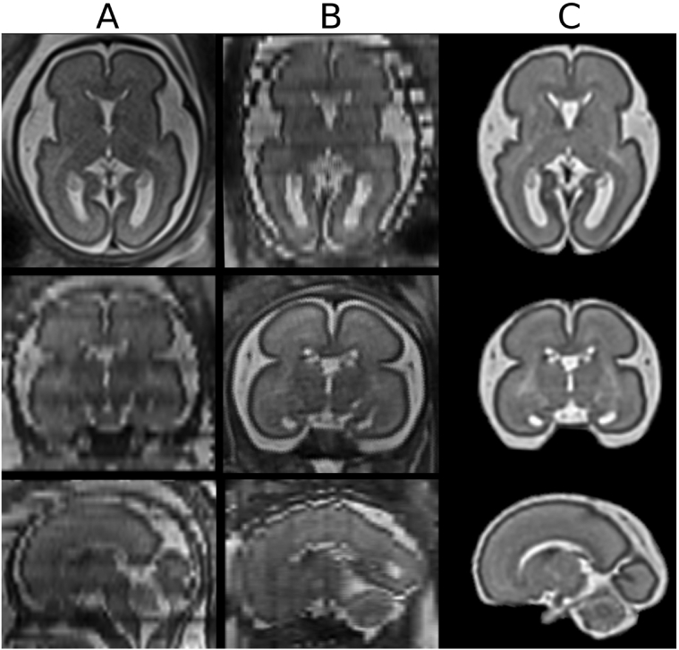
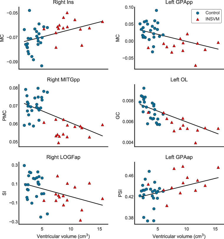
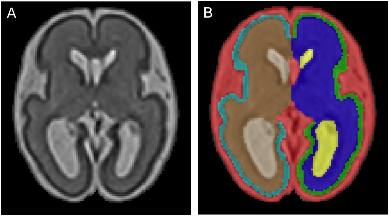
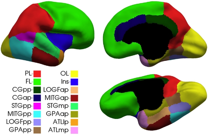
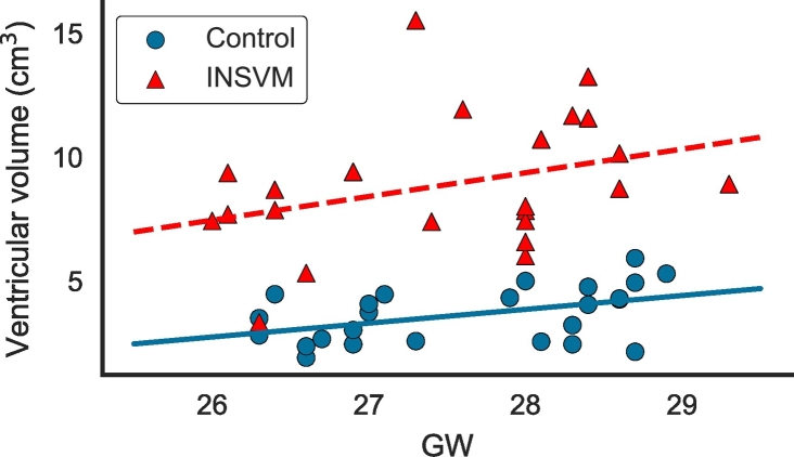
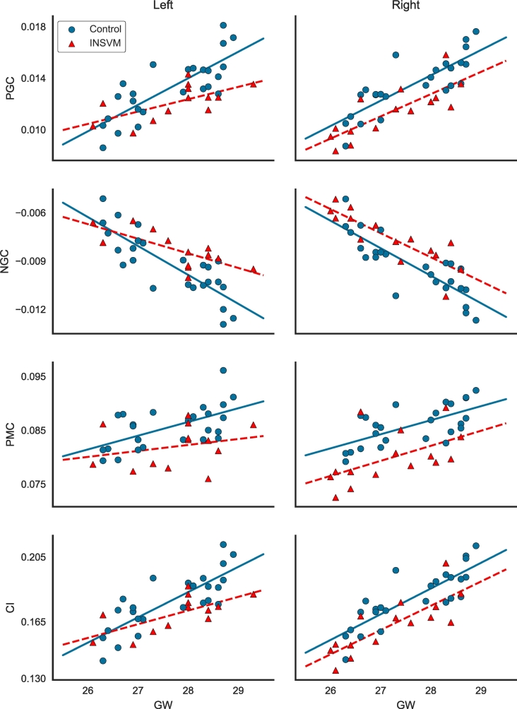
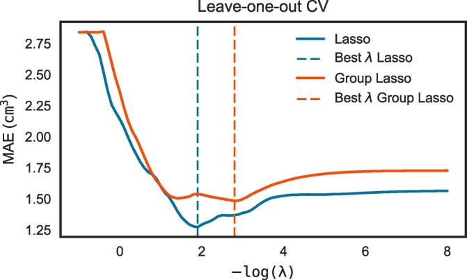
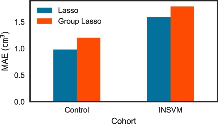
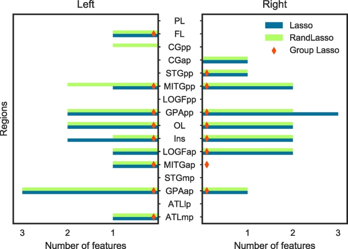
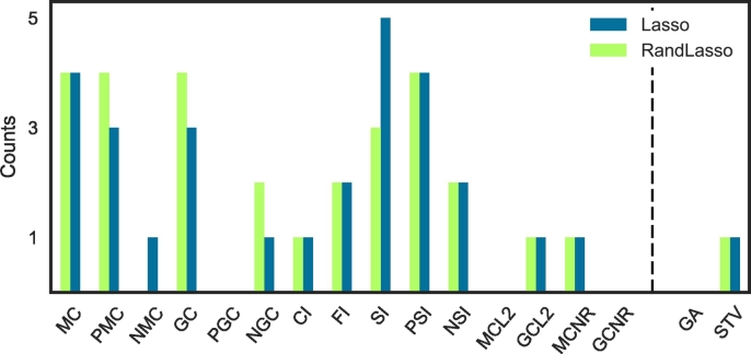
Neuroimaging of brain diseases plays a crucial role in understanding brain abnormalities and early diagnosis. Of great importance is the study of brain abnormalities and the assessment of deviations in case of maldevelopment. In this work, brain magnetic resonance images from 23 isolated non-severe ventriculomegaly (INSVM) fetuses and 25 healthy controls between 26 and 29 gestational weeks were used to identify INSVM-related cortical folding deviations from normative development. Since these alterations may reflect abnormal neurodevelopment, our working hypothesis is that markers of cortical folding can provide cues to improve the prediction of later neurodevelopmental problems in INSVM subjects. We analyzed the relationship of ventricular enlargement with cortical folding alterations in a regional basis using several curvature-based measures describing the folding of each cortical region. Statistical analysis (global and hemispheric) and sparse linear regression approaches were then used to find the cortical regions whose folding is associated with ventricular dilation. Results from both approaches were in great accordance, showing a significant cortical folding decrease in the insula, posterior part of the temporal lobe and occipital lobe. Moreover, compared to the global analysis, stronger ipsilateral associations of ventricular enlargement with reduced cortical folding were encountered by the hemispheric analysis. Our findings confirm and extend previous studies by identifying various cortical regions and emphasizing ipsilateral effects of ventricular enlargement in altered folding. This suggests that INSVM is an indicator of altered cortical development, and moreover, cortical regions with reduced folding constitute potential prognostic biomarkers to be used in follow-up studies to decipher the outcome of INSVM fetuses.

摘要

脑疾病的神经影像学在理解脑异常和早期诊断中起着至关重要的作用。研究脑异常和评估发育不良时的偏差非常重要。在这项工作中,我们使用了 23 例孤立性非严重脑室扩大(INSVM)胎儿和 25 例 26 至 29 孕周的健康对照的脑磁共振图像,以确定 INSVM 相关的皮质折叠偏离正常发育的情况。由于这些改变可能反映了异常的神经发育,我们的工作假设是皮质折叠的标志物可以提供线索,以提高对 INSVM 受试者后期神经发育问题的预测。我们使用了几种基于曲率的测量方法,从区域基础上分析了脑室扩大与皮质折叠改变的关系,这些方法描述了每个皮质区域的折叠情况。然后,我们使用了统计分析(全局和半球)和稀疏线性回归方法来找到与脑室扩张相关的折叠皮质区域。这两种方法的结果非常一致,表明在脑岛、颞叶后部和枕叶皮质折叠明显减少。此外,与全局分析相比,半球分析发现脑室扩大与皮质折叠减少之间存在更强的同侧关联。我们的发现通过确定各种皮质区域并强调脑室扩大对折叠的同侧影响,证实并扩展了之前的研究。这表明 INSVM 是皮质发育改变的一个指标,而且折叠减少的皮质区域构成了潜在的预后生物标志物,可以在后续研究中使用,以解读 INSVM 胎儿的结局。

https://cdn.ncbi.nlm.nih.gov/pmc/blobs/319f/5790022/9e6708b1653b/gr10.jpg
https://cdn.ncbi.nlm.nih.gov/pmc/blobs/319f/5790022/aceeff4a7838/gr1.jpg
https://cdn.ncbi.nlm.nih.gov/pmc/blobs/319f/5790022/cf5f451d2f66/gr2.jpg
https://cdn.ncbi.nlm.nih.gov/pmc/blobs/319f/5790022/19bd4a6a2fc4/gr3.jpg
https://cdn.ncbi.nlm.nih.gov/pmc/blobs/319f/5790022/56d2823c1d2f/gr4.jpg
https://cdn.ncbi.nlm.nih.gov/pmc/blobs/319f/5790022/98e1ed20bed8/gr5.jpg
https://cdn.ncbi.nlm.nih.gov/pmc/blobs/319f/5790022/c77b121ef5b7/gr6.jpg
https://cdn.ncbi.nlm.nih.gov/pmc/blobs/319f/5790022/6db645e63fa4/gr7.jpg
https://cdn.ncbi.nlm.nih.gov/pmc/blobs/319f/5790022/fa60cb650dd9/gr8.jpg
https://cdn.ncbi.nlm.nih.gov/pmc/blobs/319f/5790022/6c0b97f20e4f/gr9.jpg
https://cdn.ncbi.nlm.nih.gov/pmc/blobs/319f/5790022/9e6708b1653b/gr10.jpg

相似文献

1
Cortical folding alterations in fetuses with isolated non-severe ventriculomegaly.孤立性非重度脑室扩张胎儿的皮质折叠改变。
Neuroimage Clin. 2018 Jan 28;18:103-114. doi: 10.1016/j.nicl.2018.01.006. eCollection 2018.
2
Global and Regional Changes in Cortical Development Assessed by MRI in Fetuses with Isolated Nonsevere Ventriculomegaly Correlate with Neonatal Neurobehavior.MRI 评估孤立性非重度脑室扩张胎儿皮质发育的全球和区域性变化与新生儿神经行为相关。
AJNR Am J Neuroradiol. 2019 Sep;40(9):1567-1574. doi: 10.3174/ajnr.A6165.
3
Altered cortical development in fetuses with isolated nonsevere ventriculomegaly assessed by neurosonography.神经超声评估孤立性非重度脑室扩张胎儿的皮质发育改变。
Prenat Diagn. 2018 Apr;38(5):365-375. doi: 10.1002/pd.5240.
4
Learning to combine complementary segmentation methods for fetal and 6-month infant brain MRI segmentation.学习将互补的分割方法结合用于胎儿和 6 个月大婴儿的脑 MRI 分割。
Comput Med Imaging Graph. 2018 Nov;69:52-59. doi: 10.1016/j.compmedimag.2018.08.007. Epub 2018 Aug 28.
5
A novel approach to multiple anatomical shape analysis: Application to fetal ventriculomegaly.一种用于多种解剖形状分析的新方法:应用于胎儿脑室扩大。
Med Image Anal. 2020 Aug;64:101750. doi: 10.1016/j.media.2020.101750. Epub 2020 Jun 10.
6
Role of magnetic resonance imaging in fetuses with mild or moderate ventriculomegaly in the era of fetal neurosonography: systematic review and meta-analysis.磁共振成像在胎儿神经超声时代轻度或中度脑室扩张胎儿中的作用:系统评价和荟萃分析。
Ultrasound Obstet Gynecol. 2019 Aug;54(2):164-171. doi: 10.1002/uog.20197. Epub 2019 Jul 11.
7
Asymmetric cortical development and prognosis in fetuses with isolated mild fetal ventriculomegaly: an observational prospective study.孤立性轻度胎儿脑室扩大胎儿的不对称皮质发育与预后:一项前瞻性观察研究。
BMC Pregnancy Childbirth. 2021 Mar 10;21(1):199. doi: 10.1186/s12884-021-03692-x.
8
Volumetric and surface-based 3D MRI analyses of fetal isolated mild ventriculomegaly: brain morphometry in ventriculomegaly.胎儿孤立性轻度脑室扩张的容积和基于表面的 3D MRI 分析:脑室扩张中的脑形态计量学。
Brain Struct Funct. 2013 May;218(3):645-55. doi: 10.1007/s00429-012-0418-1. Epub 2012 May 1.
9
Regional Alterations in Cortical Sulcal Depth in Living Fetuses with Down Syndrome.唐氏综合征活胎大脑皮层脑沟深度的区域性变化。
Cereb Cortex. 2021 Jan 5;31(2):757-767. doi: 10.1093/cercor/bhaa255.
10
Cortical overgrowth in fetuses with isolated ventriculomegaly.孤立性脑室扩大胎儿的皮质过度生长。
Cereb Cortex. 2014 Aug;24(8):2141-50. doi: 10.1093/cercor/bht062. Epub 2013 Mar 18.

引用本文的文献

1
A simple and quantitative ultrasonographic screening method for fetal Sylvian fissure abnormalities.一种用于胎儿大脑外侧裂异常的简单定量超声筛查方法。
Eur J Med Res. 2025 Jun 18;30(1):488. doi: 10.1186/s40001-025-02708-y.
2
Longitudinal Assessment of Abnormal Cortical Folding in Fetuses and Neonates With Isolated Non-Severe Ventriculomegaly.孤立性非重度脑室扩大胎儿及新生儿皮质折叠异常的纵向评估
Brain Behav. 2025 Jan;15(1):e70255. doi: 10.1002/brb3.70255.
3
Deep cascaded registration and weakly-supervised segmentation of fetal brain MRI.

本文引用的文献

1
Toward the automatic quantification of in utero brain development in 3D structural MRI: A review.迈向三维结构磁共振成像中子宫内脑发育的自动量化:综述
Hum Brain Mapp. 2017 May;38(5):2772-2787. doi: 10.1002/hbm.23536. Epub 2017 Feb 14.
2
Perinatal and long-term outcomes in fetuses diagnosed with isolated unilateral ventriculomegaly: systematic review and meta-analysis.孤立性单侧脑室扩大胎儿的围产期及长期结局:系统评价与荟萃分析
Ultrasound Obstet Gynecol. 2017 Apr;49(4):450-459. doi: 10.1002/uog.15943. Epub 2017 Feb 28.
3
Cerebral cortex expansion and folding: what have we learned?
胎儿脑磁共振成像的深度级联配准与弱监督分割
Heliyon. 2024 Nov 19;11(1):e40148. doi: 10.1016/j.heliyon.2024.e40148. eCollection 2025 Jan 15.
4
Current Diagnostic, Counseling, and Treatment Options in Non-Severe and Severe Apparently Isolated Fetal Ventriculomegaly.非重度和重度明显孤立性胎儿脑室扩大的当前诊断、咨询及治疗选择
Biomedicines. 2024 Dec 23;12(12):2929. doi: 10.3390/biomedicines12122929.
5
T* relaxometry of fetal brain structures using low-field (0.55T) MRI.使用低场(0.55T)MRI对胎儿脑结构进行T* 弛豫测量法。
Magn Reson Med. 2025 May;93(5):1942-1953. doi: 10.1002/mrm.30409. Epub 2024 Dec 31.
6
Fetal gestational age prediction via shape descriptors of cortical development.通过皮质发育的形状描述符预测胎儿孕周。
Front Pediatr. 2024 Nov 20;12:1471080. doi: 10.3389/fped.2024.1471080. eCollection 2024.
7
Isolated Fetal Ventriculomegaly: Diagnosis and Treatment in the Prenatal Period.孤立性胎儿脑室扩大:产前诊断与治疗
Children (Basel). 2024 Aug 8;11(8):957. doi: 10.3390/children11080957.
8
Volume development changes in the occipital lobe gyrus assessed by MRI in fetuses with isolated ventriculomegaly correlate with neurological development in infancy and early childhood.MRI 评估孤立性脑室扩大胎儿枕叶脑回体积发育变化与婴儿期和幼儿期神经发育相关。
J Perinatol. 2024 Aug;44(8):1178-1185. doi: 10.1038/s41372-024-02012-3. Epub 2024 May 27.
9
Prenatal diagnosis and pregnancy outcomes in fetuses with ventriculomegaly.脑室扩大胎儿的产前诊断与妊娠结局
Front Med (Lausanne). 2024 May 9;11:1349171. doi: 10.3389/fmed.2024.1349171. eCollection 2024.
10
Fetal neuroimaging applications for diagnosis and counseling of brain anomalies: Current practice and future diagnostic strategies.胎儿神经影像学在脑畸形的诊断和咨询中的应用:当前实践和未来的诊断策略。
Semin Fetal Neonatal Med. 2024 Feb;29(1):101525. doi: 10.1016/j.siny.2024.101525. Epub 2024 Apr 10.
大脑皮层的扩张与折叠:我们学到了什么?
EMBO J. 2016 May 17;35(10):1021-44. doi: 10.15252/embj.201593701. Epub 2016 Apr 7.
4
Comparison of cortical folding measures for evaluation of developing human brain.用于评估发育中人类大脑的皮质折叠测量方法的比较
Neuroimage. 2016 Jan 15;125:780-790. doi: 10.1016/j.neuroimage.2015.11.001. Epub 2015 Nov 6.
5
Cortical folding and the potential for prognostic neuroimaging in schizophrenia.精神分裂症中的皮质折叠及预后神经影像学潜力
Br J Psychiatry. 2015 Nov;207(5):458-9. doi: 10.1192/bjp.bp.114.155796. Epub 2015 Jul 23.
6
Morphological abnormalities in prefrontal surface area and thalamic volume in attention deficit/hyperactivity disorder.注意缺陷多动障碍患者前额叶表面积和丘脑体积的形态学异常
Psychiatry Res. 2015 Aug 30;233(2):225-32. doi: 10.1016/j.pscychresns.2015.07.004. Epub 2015 Jul 7.
7
Development of Cortical Morphology Evaluated with Longitudinal MR Brain Images of Preterm Infants.利用早产儿脑部纵向磁共振图像评估皮质形态的发育
PLoS One. 2015 Jul 10;10(7):e0131552. doi: 10.1371/journal.pone.0131552. eCollection 2015.
8
Construction of a fetal spatio-temporal cortical surface atlas from in utero MRI: Application of spectral surface matching.利用宫内磁共振成像构建胎儿时空皮质表面图谱:谱表面匹配的应用
Neuroimage. 2015 Oct 15;120:467-80. doi: 10.1016/j.neuroimage.2015.05.087. Epub 2015 Jun 10.
9
Assessment of MRI-Based Automated Fetal Cerebral Cortical Folding Measures in Prediction of Gestational Age in the Third Trimester.基于磁共振成像的胎儿大脑皮质折叠自动测量在孕晚期孕周预测中的评估
AJNR Am J Neuroradiol. 2015 Jul;36(7):1369-74. doi: 10.3174/ajnr.A4357. Epub 2015 Jun 4.
10
Toward Probabilistic Diagnosis and Understanding of Depression Based on Functional MRI Data Analysis with Logistic Group LASSO.基于逻辑组套索功能磁共振成像数据分析的抑郁症概率诊断与理解
PLoS One. 2015 May 1;10(5):e0123524. doi: 10.1371/journal.pone.0123524. eCollection 2015.