Chen Yu-Chen, Liu Shenghua, Lv Han, Bo Fan, Feng Yuan, Chen Huiyou, Xu Jin-Jing, Yin Xindao, Wang Shukui, Gu Jian-Ping
Department of Radiology, Nanjing First Hospital, Nanjing Medical University, Nanjing, China.
Department of Radiology, Beijing Friendship Hospital, Capital Medical University, Beijing, China.
Front Neurosci. 2018 Jan 23;12:9. doi: 10.3389/fnins.2018.00009. eCollection 2018.
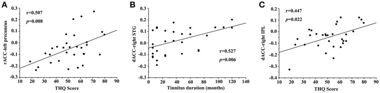
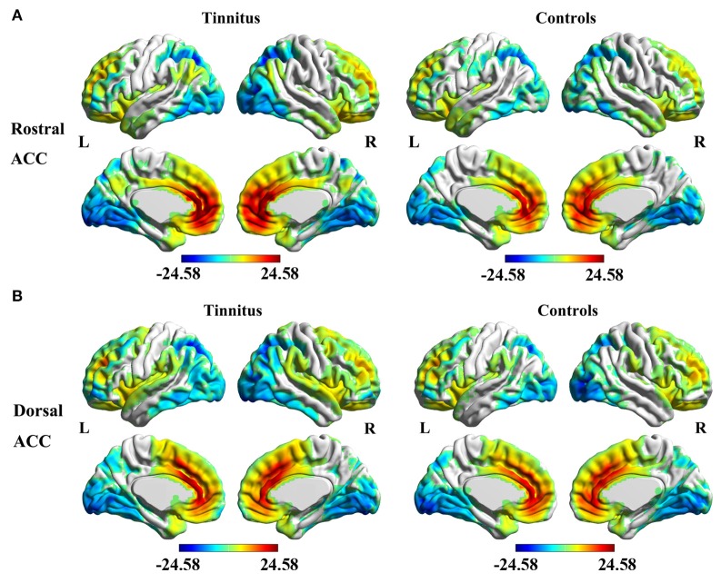
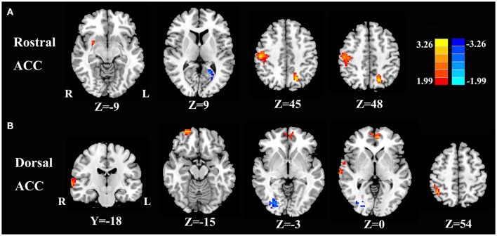
The anterior cingulate cortex (ACC) has been suggested to be involved in chronic subjective tinnitus. Tinnitus may arise from aberrant functional coupling between the ACC and cerebral cortex. To explore this hypothesis, we used resting-state functional magnetic resonance imaging (fMRI) to illuminate the functional connectivity (FC) network of the ACC subregions in chronic tinnitus patients. Resting-state fMRI scans were obtained from 31 chronic right-sided tinnitus patients and 40 healthy controls (age, sex, and education well-matched) in this study. Rostral ACC and dorsal ACC were selected as seed regions to investigate the intrinsic FC with the whole brain. The resulting FC patterns were correlated with clinical tinnitus characteristics including the tinnitus duration and tinnitus distress. Compared with healthy controls, chronic tinnitus patients showed disrupted FC patterns of ACC within several brain networks, including the auditory cortex, prefrontal cortex, visual cortex, and default mode network (DMN). The Tinnitus Handicap Questionnaires (THQ) scores showed positive correlations with increased FC between the rostral ACC and left precuneus ( = 0.507, = 0.008) as well as the dorsal ACC and right inferior parietal lobe ( = 0.447, = 0.022). Chronic tinnitus patients have abnormal FC networks originating from ACC to other selected brain regions that are associated with specific tinnitus characteristics. Resting-state ACC-cortical FC disturbances may play an important role in neuropathological features underlying chronic tinnitus.
前扣带回皮质(ACC)被认为与慢性主观性耳鸣有关。耳鸣可能源于ACC与大脑皮质之间异常的功能耦合。为了探究这一假设,我们使用静息态功能磁共振成像(fMRI)来阐明慢性耳鸣患者ACC亚区域的功能连接(FC)网络。在本研究中,从31名慢性右侧耳鸣患者和40名健康对照者(年龄、性别和教育程度匹配良好)中获取了静息态fMRI扫描数据。选择喙状ACC和背侧ACC作为种子区域,以研究其与全脑的内在FC。将得到的FC模式与包括耳鸣持续时间和耳鸣困扰在内的临床耳鸣特征进行相关性分析。与健康对照者相比,慢性耳鸣患者在包括听觉皮质、前额叶皮质、视觉皮质和默认模式网络(DMN)在内的几个脑网络中显示出ACC的FC模式中断。耳鸣障碍问卷(THQ)评分与喙状ACC和左侧楔前叶之间(r = 0.507,p = 0.008)以及背侧ACC和右侧顶下小叶之间(r = 0.447,p = 0.022)的FC增加呈正相关。慢性耳鸣患者存在源自ACC至其他选定脑区的异常FC网络,这些网络与特定的耳鸣特征相关。静息态ACC - 皮质FC紊乱可能在慢性耳鸣潜在的神经病理特征中起重要作用。