• 文献检索
  • 文档翻译
  • 深度研究
  • 学术资讯
  • Suppr Zotero 插件Zotero 插件
  • 邀请有礼
  • 套餐&价格
  • 历史记录
应用&插件
Suppr Zotero 插件Zotero 插件浏览器插件Mac 客户端Windows 客户端微信小程序
定价
高级版会员购买积分包购买API积分包
服务
文献检索文档翻译深度研究API 文档MCP 服务
关于我们
关于 Suppr公司介绍联系我们用户协议隐私条款
关注我们

Suppr 超能文献

核心技术专利:CN118964589B侵权必究
粤ICP备2023148730 号-1Suppr @ 2026

文献检索

告别复杂PubMed语法,用中文像聊天一样搜索,搜遍4000万医学文献。AI智能推荐,让科研检索更轻松。

立即免费搜索

文件翻译

保留排版,准确专业,支持PDF/Word/PPT等文件格式,支持 12+语言互译。

免费翻译文档

深度研究

AI帮你快速写综述,25分钟生成高质量综述,智能提取关键信息,辅助科研写作。

立即免费体验

并非所有的假阳性诊断都是等同的:关于在乳腺 MRI 与乳腺钼靶/数字断层合成筛查中做出的假阳性诊断的预后意义。

Not all false positive diagnoses are equal: On the prognostic implications of false-positive diagnoses made in breast MRI versus in mammography / digital tomosynthesis screening.

机构信息

Department of Diagnostic and Interventional Radiology, Hospital of the University of Aachen, RWTH, Pauwelsstrasse 30, 52074, Aachen, Germany.

Department of Pathology, Hospital of the University of Aachen, RWTH, Aachen, Germany.

出版信息

Breast Cancer Res. 2018 Feb 9;20(1):13. doi: 10.1186/s13058-018-0937-7.

DOI:10.1186/s13058-018-0937-7
PMID:29426360
原文链接:https://pmc.ncbi.nlm.nih.gov/articles/PMC5807753/
Abstract

BACKGROUND

Breast magnetic resonance imaging (MRI) has been reported to frequently result in false-positive diagnoses, limiting its positive predictive value (PPV). However, for PPV calculation, all nonmalignant tissue changes are equally considered false-positive, although the respective prognostic importance, and thus patient management implications, of different pathologies may well differ. We investigated the pathology of false-positive diagnoses made by MRI compared with radiographic (digital mammography/tomosynthesis [DM/DBT]) screening.

METHODS

We conducted an institutional review board-approved prospective analysis of 710 consecutive asymptomatic women at average risk for breast cancer who underwent vacuum biopsy with or without surgical biopsy for screen-detected DM/DBT (n = 344) or MRI (n = 366) findings. We compared the frequency of false-positive biopsies (given by PPV3), as well as the types of nonmalignant tissue changes that caused the respective false-positive biopsies. In an order of increasing relative risk of subsequent breast cancer, pathologies of false-positive biopsies were categorized as nonproliferative, simple proliferative, complex proliferative, or atypical proliferative (including lobular carcinoma in situ/lobular intraepithelial neoplasia). The Mann-Whitney U test was used to compare distributions.

RESULTS

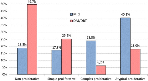
Histology yielded nonmalignant tissue in 202 of 366 biopsies done for positive MRI studies and 195 of 344 biopsies for positive DM/DBT studies, respectively, yielding a similar PPV3 percentages of 44.8% (164 of 202) and 43.3% (149 of 202) for both methods. However, the distribution of tissue types that caused false-positive diagnoses differed significantly (p < 0.0001). On the basis of MRI, high-risk atypical proliferative changes (40.1%; 81 of 202) were most common, followed by complex proliferative changes (23.8%; 48 of 202). In DM/DBT, low-risk, nonproliferative changes were the dominant reason for false-positive diagnoses (49.7%; 97 of 195), followed by simple proliferative changes (25.2%; 51 of 195). Low-risk nonproliferative changes resulted in false-positive diagnoses based on MRI as infrequently as did high-risk atypical proliferative changes based on DM/DBT (18.8% [38 of 202] vs. 18.0% [35 of 195]). The likelihood of a false-positive diagnosis including atypias was twice as high in women undergoing biopsy for MRI findings (81 of 202; 40%) as for those with DM/DBT findings (35 of 195; 18%).

CONCLUSIONS

The prognostic importance, and thus the clinical implications, of false-positive diagnoses made on the basis of breast MRI vs. radiographic screening differed significantly, with a reversed prevalence of high- and low-risk lesions. This should be taken into account when discussing the rate of false-positive diagnoses (i.e., PPV levels of MRI vs. radiographic screening). Current benchmarks that rate the utility of breast cancer screening programs (i.e., cancer detection rates and PPVs) do not reflect these substantial biological differences and the different prognostic implications.

https://cdn.ncbi.nlm.nih.gov/pmc/blobs/097d/5807753/b15598c21ca1/13058_2018_937_Fig3_HTML.jpg
https://cdn.ncbi.nlm.nih.gov/pmc/blobs/097d/5807753/d05a3805f394/13058_2018_937_Fig1_HTML.jpg
https://cdn.ncbi.nlm.nih.gov/pmc/blobs/097d/5807753/f64137809a9e/13058_2018_937_Fig2_HTML.jpg
https://cdn.ncbi.nlm.nih.gov/pmc/blobs/097d/5807753/b15598c21ca1/13058_2018_937_Fig3_HTML.jpg
https://cdn.ncbi.nlm.nih.gov/pmc/blobs/097d/5807753/d05a3805f394/13058_2018_937_Fig1_HTML.jpg
https://cdn.ncbi.nlm.nih.gov/pmc/blobs/097d/5807753/f64137809a9e/13058_2018_937_Fig2_HTML.jpg
https://cdn.ncbi.nlm.nih.gov/pmc/blobs/097d/5807753/b15598c21ca1/13058_2018_937_Fig3_HTML.jpg
摘要

背景

据报道,乳腺磁共振成像(MRI)经常导致假阳性诊断,从而限制了其阳性预测值(PPV)。然而,对于 PPV 计算,所有非恶性组织变化都被同等视为假阳性,尽管不同病理的各自预后重要性,以及因此对患者管理的影响,可能存在差异。我们研究了 MRI 筛查与放射学(数字乳腺 X 线摄影/断层合成[DM/DBT])筛查相比假阳性诊断的病理学。

方法

我们对平均有乳腺癌风险的 710 例连续无症状女性进行了机构审查委员会批准的前瞻性分析,这些女性接受了真空活检,或者根据 DM/DBT(n=344)或 MRI(n=366)筛查结果进行了手术活检。我们比较了假阳性活检(由 PPV3 给出)的频率,以及导致各自假阳性活检的非恶性组织变化的类型。根据随后发生乳腺癌的相对风险递增顺序,假阳性活检的病理学被归类为非增生性、单纯增生性、复杂增生性或非典型增生性(包括小叶原位癌/小叶上皮内瘤变)。采用 Mann-Whitney U 检验比较分布情况。

结果

在 MRI 阳性研究的 366 例活检中,组织学显示非恶性组织的有 202 例,在 DM/DBT 阳性研究的 344 例活检中,组织学显示非恶性组织的有 195 例,分别产生了 44.8%(202 例中有 164 例)和 43.3%(202 例中有 149 例)的相似 PPV3 百分比。然而,导致假阳性诊断的组织类型分布差异显著(p<0.0001)。基于 MRI,高风险非典型增生性改变(40.1%;202 例中有 81 例)最为常见,其次是复杂增生性改变(23.8%;202 例中有 48 例)。在 DM/DBT 中,低风险、非增生性改变是假阳性诊断的主要原因(49.7%;195 例中有 97 例),其次是简单增生性改变(25.2%;195 例中有 51 例)。低风险非增生性改变导致假阳性诊断的频率与高风险非典型增生性改变导致基于 DM/DBT 的假阳性诊断的频率一样低(18.8%[202 例中有 38 例] vs. 18.0%[195 例中有 35 例])。MRI 活检的假阳性诊断中包括非典型增生的可能性是 DM/DBT 活检的两倍(81 例中有 40%[202 例],而 35 例中有 18%[195 例])。

结论

MRI 与放射学筛查相比,假阳性诊断的预后重要性,因此临床意义存在显著差异,高风险和低风险病变的发生率也存在反转。在讨论假阳性诊断率(即 MRI 与放射学筛查的 PPV 水平)时,应考虑到这一点。当前评估乳腺癌筛查计划效用的基准(即癌症检出率和 PPV)并没有反映这些实质性的生物学差异和不同的预后影响。

相似文献

1
Not all false positive diagnoses are equal: On the prognostic implications of false-positive diagnoses made in breast MRI versus in mammography / digital tomosynthesis screening.并非所有的假阳性诊断都是等同的:关于在乳腺 MRI 与乳腺钼靶/数字断层合成筛查中做出的假阳性诊断的预后意义。
Breast Cancer Res. 2018 Feb 9;20(1):13. doi: 10.1186/s13058-018-0937-7.
2
3
4
Detection Rates for Benign and Malignant Diagnoses on Breast Cancer Screening With Digital Breast Tomosynthesis in a Statewide Mammography Registry Study.在全州乳腺摄影登记研究中,数字乳腺断层合成摄影术用于乳腺癌筛查的良性和恶性诊断检出率。
AJR Am J Roentgenol. 2019 Mar;212(3):706-711. doi: 10.2214/AJR.18.20255. Epub 2019 Jan 23.
5
Effectiveness of Digital Breast Tomosynthesis Compared With Digital Mammography: Outcomes Analysis From 3 Years of Breast Cancer Screening.数字乳腺断层合成摄影与数字乳腺钼靶摄影的有效性比较:3 年乳腺癌筛查的结果分析。
JAMA Oncol. 2016 Jun 1;2(6):737-43. doi: 10.1001/jamaoncol.2015.5536.
6
Assessment of disease extent on contrast-enhanced MRI in breast cancer detected at digital breast tomosynthesis versus digital mammography alone.在数字乳腺断层合成与单纯数字乳腺摄影检测到的乳腺癌中,通过对比增强磁共振成像评估疾病范围。
Clin Radiol. 2017 Jul;72(7):573-579. doi: 10.1016/j.crad.2017.02.013. Epub 2017 Mar 17.
7
Comparison of performance metrics with digital 2D versus tomosynthesis mammography in the diagnostic setting.在诊断环境中比较数字二维与断层合成乳腺摄影的性能指标。
Eur Radiol. 2019 Feb;29(2):477-484. doi: 10.1007/s00330-018-5596-7. Epub 2018 Jul 2.
8
Digital breast tomosynthesis plus mammography, magnetic resonance imaging plus mammography and mammography alone: A comparison of diagnostic performance in symptomatic women.数字乳腺断层合成联合乳腺X线摄影、磁共振成像联合乳腺X线摄影及单纯乳腺X线摄影:有症状女性诊断性能的比较
Clin Hemorheol Microcirc. 2017;66(2):105-116. doi: 10.3233/CH-16242.
9
Digital Mammography versus Digital Mammography Plus Tomosynthesis for Breast Cancer Screening: The Reggio Emilia Tomosynthesis Randomized Trial.数字乳腺 X 线摄影与数字乳腺 X 线摄影加断层合成技术用于乳腺癌筛查的比较:雷焦艾米利亚断层合成随机试验。
Radiology. 2018 Aug;288(2):375-385. doi: 10.1148/radiol.2018172119. Epub 2018 Jun 5.
10
Multicenter Evaluation of Breast Cancer Screening with Digital Breast Tomosynthesis in Combination with Synthetic versus Digital Mammography.多中心研究:数字乳腺断层合成摄影与数字乳腺钼靶摄影联合用于乳腺癌筛查的评估。
Radiology. 2020 Dec;297(3):545-553. doi: 10.1148/radiol.2020200240. Epub 2020 Oct 13.

引用本文的文献

1
Development of uMUC-1 Targeted NEMO Particles with pH-Activatable MRI Signals for Enhanced Detection of Malignant Breast Cancer Cells.具有pH可激活MRI信号的uMUC-1靶向NEMO颗粒用于增强检测恶性乳腺癌细胞的研究进展。
ACS Appl Bio Mater. 2025 May 19;8(5):4251-4261. doi: 10.1021/acsabm.5c00365. Epub 2025 May 1.
2
Optimizing breast MRI diagnosis: the Kaiser Score's impact on reducing unnecessary biopsies.优化乳腺磁共振成像诊断:凯泽评分对减少不必要活检的影响。
Eur Radiol. 2025 Mar;35(3):1502-1503. doi: 10.1007/s00330-024-11325-y. Epub 2025 Jan 2.
3
Machine learning radiomics based on intra and peri tumor PA/US images distinguish between luminal and non-luminal tumors in breast cancers.

本文引用的文献

1
Abbreviated breast MRI for screening women with dense breast: the EA1141 trial.用于筛查乳腺致密女性的简化乳腺磁共振成像:EA1141试验
Br J Radiol. 2018 Oct;91(1090):20170441. doi: 10.1259/bjr.20170441. Epub 2017 Oct 27.
2
Performance Benchmarks for Screening Breast MR Imaging in Community Practice.社区实践中乳腺磁共振成像筛查的性能基准
Radiology. 2017 Oct;285(1):44-52. doi: 10.1148/radiol.2017162033. Epub 2017 Jun 5.
3
Supplemental Breast MR Imaging Screening of Women with Average Risk of Breast Cancer.补充乳腺磁共振成像筛查对乳腺癌平均风险女性。
基于肿瘤内部及周边PA/US图像的机器学习放射组学可区分乳腺癌中的管腔型肿瘤和非管腔型肿瘤。
Photoacoustics. 2024 Sep 23;40:100653. doi: 10.1016/j.pacs.2024.100653. eCollection 2024 Dec.
4
The Role of MRI in Breast Cancer and Breast Conservation Therapy.磁共振成像在乳腺癌及保乳治疗中的作用
Cancers (Basel). 2024 Jun 1;16(11):2122. doi: 10.3390/cancers16112122.
5
Application value of MRI-guided wire localization to the non-palpable breast lesions only shown in Breast MRI.MRI引导下金属丝定位对仅在乳腺MRI中显示的不可触及乳腺病变的应用价值。
Front Oncol. 2024 May 24;14:1325362. doi: 10.3389/fonc.2024.1325362. eCollection 2024.
6
Breast density knowledge and willingness to delay treatment for pre-operative breast cancer imaging among women with a personal history of breast cancer.有乳腺癌个人病史的女性对乳房密度的认知和对术前乳腺癌影像学检查延迟治疗的意愿。
Breast Cancer Res. 2024 Apr 29;26(1):73. doi: 10.1186/s13058-024-01820-x.
7
Optimizing breast cancer diagnosis with photoacoustic imaging: An analysis of intratumoral and peritumoral radiomics.利用光声成像优化乳腺癌诊断:瘤内和瘤周放射组学分析
Photoacoustics. 2024 Apr 9;38:100606. doi: 10.1016/j.pacs.2024.100606. eCollection 2024 Aug.
8
Immediate and delayed risk of breast cancer associated with classic lobular carcinoma in situ and its variants.经典型乳腺小叶原位癌及其变异型与乳腺癌的即刻和延迟风险。
Breast Cancer Res Treat. 2024 Jun;205(3):545-554. doi: 10.1007/s10549-024-07261-6. Epub 2024 Mar 12.
9
Improving signal-to-noise ratio by maximal convolution of longitudinal and transverse magnetization components in MRI: application to the breast cancer detection.通过在 MRI 中对纵向和横向磁化分量进行最大卷积来提高信噪比:在乳腺癌检测中的应用。
Med Biol Eng Comput. 2024 Mar;62(3):941-954. doi: 10.1007/s11517-023-02994-w. Epub 2023 Dec 15.
10
How Does Diagnostic Accuracy Evolve with Increased Breast MRI Experience?随着乳腺 MRI 经验的增加,诊断准确性如何演变?
Tomography. 2023 Nov 6;9(6):2067-2078. doi: 10.3390/tomography9060162.
Radiology. 2017 May;283(2):361-370. doi: 10.1148/radiol.2016161444. Epub 2017 Feb 21.
4
Impact of a False-Positive Screening Mammogram on Subsequent Screening Behavior and Stage at Breast Cancer Diagnosis.乳腺钼靶筛查假阳性对后续筛查行为及乳腺癌诊断分期的影响。
Cancer Epidemiol Biomarkers Prev. 2017 Mar;26(3):397-403. doi: 10.1158/1055-9965.EPI-16-0524. Epub 2017 Feb 9.
5
Magnetic Resonance Imaging as an Adjunct to Mammography for Breast Cancer Screening in Women at Less Than High Risk for Breast Cancer: A Health Technology Assessment.磁共振成像作为乳腺钼靶检查的辅助手段用于乳腺癌低风险女性的乳腺癌筛查:一项卫生技术评估。
Ont Health Technol Assess Ser. 2016 Nov 1;16(20):1-30. eCollection 2016.
6
Compliance With Screening Mammography Guidelines After a False-Positive Mammogram.乳腺钼靶检查结果为假阳性后对乳腺钼靶筛查指南的依从性。
J Am Coll Radiol. 2016 Sep;13(9):1032-8. doi: 10.1016/j.jacr.2016.03.016. Epub 2016 May 24.
7
Breast Cancers Detected at Screening MR Imaging and Mammography in Patients at High Risk: Method of Detection Reflects Tumor Histopathologic Results.高危患者筛查性磁共振成像和乳腺钼靶检查发现的乳腺癌:检测方法反映肿瘤组织病理学结果。
Radiology. 2016 Sep;280(3):716-22. doi: 10.1148/radiol.2016151419. Epub 2016 Apr 20.
8
Treatment and Long-Term Risks for Patients With a Diagnosis of Ductal Carcinoma In Situ.导管原位癌患者的治疗及长期风险
JAMA Oncol. 2016 Mar;2(3):397-8. doi: 10.1001/jamaoncol.2015.4860.
9
Risk of breast cancer after false-positive results in mammographic screening.乳腺钼靶筛查假阳性结果后患乳腺癌的风险。
Cancer Med. 2016 Jun;5(6):1298-306. doi: 10.1002/cam4.646. Epub 2016 Feb 25.
10
Management of atypical lobular hyperplasia, atypical ductal hyperplasia, and lobular carcinoma in situ.非典型小叶增生、非典型导管增生及小叶原位癌的管理
Expert Rev Anticancer Ther. 2016;16(3):335-46. doi: 10.1586/14737140.2016.1143362. Epub 2016 Feb 6.