• 文献检索
  • 文档翻译
  • 深度研究
  • 学术资讯
  • Suppr Zotero 插件Zotero 插件
  • 邀请有礼
  • 套餐&价格
  • 历史记录
应用&插件
Suppr Zotero 插件Zotero 插件浏览器插件Mac 客户端Windows 客户端微信小程序
定价
高级版会员购买积分包购买API积分包
服务
文献检索文档翻译深度研究API 文档MCP 服务
关于我们
关于 Suppr公司介绍联系我们用户协议隐私条款
关注我们

Suppr 超能文献

核心技术专利:CN118964589B侵权必究
粤ICP备2023148730 号-1Suppr @ 2026

文献检索

告别复杂PubMed语法,用中文像聊天一样搜索,搜遍4000万医学文献。AI智能推荐,让科研检索更轻松。

立即免费搜索

文件翻译

保留排版,准确专业,支持PDF/Word/PPT等文件格式,支持 12+语言互译。

免费翻译文档

深度研究

AI帮你快速写综述,25分钟生成高质量综述,智能提取关键信息,辅助科研写作。

立即免费体验

急性和慢性原发性闭角型青光眼(青光眼)患眼与对侧眼的差异:一项超声生物显微镜定量研究。

Differences between fellow eyes of acute and chronic primary angle closure (glaucoma): An ultrasound biomicroscopy quantitative study.

作者信息

Li Mengwei, Chen Yuhong, Chen Xiaoxiao, Zhu Wenqing, Chen Xueli, Wang Xiaolei, Fang Yuan, Kong Xiangmei, Dai Yi, Chen Junyi, Sun Xinghuai

机构信息

Department of Ophthalmology and Visual Science, Eye, Ear, Nose and Throat Hospital, Shanghai Medical College of Fudan University, Shanghai, China.

Key Laboratory of Myopia, Ministry of Health (Fudan University) and Key Laboratory of Visual Impairment and Restoration of Shanghai, Shanghai, China.

出版信息

PLoS One. 2018 Feb 15;13(2):e0193006. doi: 10.1371/journal.pone.0193006. eCollection 2018.

DOI:10.1371/journal.pone.0193006
PMID:29447247
原文链接:https://pmc.ncbi.nlm.nih.gov/articles/PMC5814014/
Abstract

PURPOSE

To compare various biometric parameters between fellow eyes of acute primary angle closure (glaucoma) [APAC(G)] and fellow eyes of chronic primary angle closure (glaucoma) [CPAC(G)].

METHODS

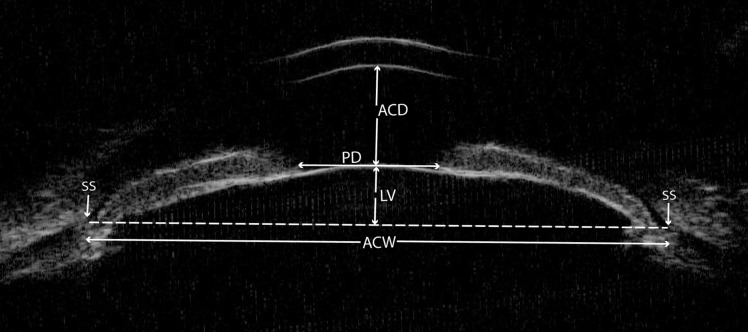
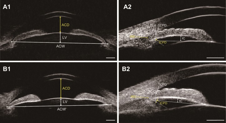
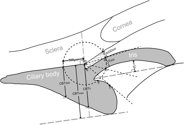
Ultrasound biomicroscopy examinations were performed on 47 patients with unilateral APAC(G) and 41 patients with asymmetric CPAC(G) before laser peripheral iridotomy and pilocarpine treatment. Anterior chamber depth and width (ACD and ACW), lens vault (LV), iris curvature (IC), iris root distance (IRD), trabecular-ciliary process distance (TCPD), iris-ciliary process distance (ICPD), trabecular-ciliary angle (TCA), and other biometric parameters were compared between fellow eyes of APAC(G) and fellow eyes of CAPC(G).

RESULTS

Compared with fellow eyes of CPAC(G), fellow eyes of APAC(G) had smaller ACD (P < 0.001), ACW (P = 0.007), TCPD (P = 0.016), ICPD (P = 0.008), and TCA (P = 0.006), as well as larger LV (P = 0.002), IC (P = 0.012), and IRD (P = 0.003). On multivariate logistic regression analyses, a 0.1 mm decrease in ACD (odds ratio [OR]: 0.705, 95%CI: 0.564-0.880, P = 0.002), ICPD (OR: 0.557, 95%CI: 0.335-0.925, P = 0.024), and a 0.1 mm increase in IRD (OR: 2.707, 95%CI: 1.025-7.149, P = 0.045), was significantly associated with occurrence of acute angle closures.

CONCLUSIONS

Fellow eyes of APAC(G) had smaller anterior segment dimensions, higher LV, more posterior iris insertion, greater IC, and more anteriorly rotated ciliary body compared with fellow eyes of CPAC(G). ACD, ICPD, and IRD were the three most important parameters that distinguish eyes predisposed to APAC(G) or CPAC(G).

摘要

目的

比较急性原发性闭角型青光眼[APAC(G)]患眼的对侧眼与慢性原发性闭角型青光眼[CPAC(G)]患眼的对侧眼之间的各种生物测量参数。

方法

对47例单侧APAC(G)患者和41例不对称CPAC(G)患者在进行激光周边虹膜切开术和毛果芸香碱治疗前进行超声生物显微镜检查。比较APAC(G)患眼的对侧眼与CAPC(G)患眼的对侧眼前房深度和宽度(ACD和ACW)、晶状体拱高(LV)、虹膜曲率(IC)、虹膜根部距离(IRD)、小梁-睫状体距离(TCPD)、虹膜-睫状体距离(ICPD)、小梁-睫状体角(TCA)及其他生物测量参数。

结果

与CPAC(G)患眼的对侧眼相比,APAC(G)患眼的对侧眼ACD更小(P<0.001)、ACW更小(P = 0.007)、TCPD更小(P = 0.016)、ICPD更小(P = 0.008)、TCA更小(P = 0.006),而LV更大(P = 0.002)、IC更大(P = 0.012)、IRD更大(P = 0.003)。多因素逻辑回归分析显示,ACD每减少0.1mm(比值比[OR]:0.705,95%可信区间[CI]:0.564 - 0.880,P = 0.002)、ICPD每减少0.1mm(OR:0.557,95%CI:0.335 - 0.925,P = 0.024)以及IRD每增加0.1mm(OR:2.707,CI:1.025 - 7.149,P = 0.045)与急性房角关闭的发生显著相关。

结论

与CPAC(G)患眼的对侧眼相比,APAC(G)患眼的对侧眼前节尺寸更小、LV更高、虹膜插入更靠后、IC更大且睫状体旋转更靠前。ACD、ICPD和IRD是区分易患APAC(G)或CPAC(G)眼的三个最重要参数。

https://cdn.ncbi.nlm.nih.gov/pmc/blobs/e1c5/5814014/c2e4f2f844a3/pone.0193006.g003.jpg
https://cdn.ncbi.nlm.nih.gov/pmc/blobs/e1c5/5814014/c87670960a73/pone.0193006.g001.jpg
https://cdn.ncbi.nlm.nih.gov/pmc/blobs/e1c5/5814014/744461ea700b/pone.0193006.g002.jpg
https://cdn.ncbi.nlm.nih.gov/pmc/blobs/e1c5/5814014/c2e4f2f844a3/pone.0193006.g003.jpg
https://cdn.ncbi.nlm.nih.gov/pmc/blobs/e1c5/5814014/c87670960a73/pone.0193006.g001.jpg
https://cdn.ncbi.nlm.nih.gov/pmc/blobs/e1c5/5814014/744461ea700b/pone.0193006.g002.jpg
https://cdn.ncbi.nlm.nih.gov/pmc/blobs/e1c5/5814014/c2e4f2f844a3/pone.0193006.g003.jpg

相似文献

1
Differences between fellow eyes of acute and chronic primary angle closure (glaucoma): An ultrasound biomicroscopy quantitative study.急性和慢性原发性闭角型青光眼(青光眼)患眼与对侧眼的差异:一项超声生物显微镜定量研究。
PLoS One. 2018 Feb 15;13(2):e0193006. doi: 10.1371/journal.pone.0193006. eCollection 2018.
2
Novel Discoveries of Anterior Segment Parameters in Fellow Eyes of Acute Primary Angle Closure and Chronic Primary Angle Closure Glaucoma.急性原发性闭角型青光眼和慢性原发性闭角型青光眼对侧眼前节参数的新发现。
Invest Ophthalmol Vis Sci. 2021 Nov 1;62(14):6. doi: 10.1167/iovs.62.14.6.
3
Ultrasound biomicroscopic features associated with angle closure in fellow eyes of acute primary angle closure after laser iridotomy.激光虹膜切开术后急性原发性闭角型青光眼对侧眼与房角关闭相关的超声生物显微镜特征
Ophthalmology. 2009 Mar;116(3):444-448.e2. doi: 10.1016/j.ophtha.2008.10.019. Epub 2009 Jan 20.
4
Quantitative Measurements of the Ciliary Body in Eyes With Acute Primary-Angle Closure.急性原发性闭角型青光眼患者睫状体的定量测量
Invest Ophthalmol Vis Sci. 2016 Jun 1;57(7):3299-305. doi: 10.1167/iovs.16-19558.
5
Difference of uveal parameters between the acute primary angle closure eyes and the fellow eyes.急性原发性闭角型青光眼患眼与对侧眼之间葡萄膜参数的差异。
Eye (Lond). 2018 Jul;32(7):1174-1182. doi: 10.1038/s41433-018-0056-9. Epub 2018 Mar 1.
6
Anterior Segment Features in Nanophthalmos With Secondary Chronic Angle Closure Glaucoma: An Ultrasound Biomicroscopy Study.超声生物显微镜研究小眼球伴发性慢性闭角型青光眼眼前节特征
Invest Ophthalmol Vis Sci. 2019 May 1;60(6):2248-2256. doi: 10.1167/iovs.19-26867.
7
Postiridotomy ultrasound biomicroscopy features in the fellow eye of Chinese patients with acute primary angle-closure and chronic primary angle-closure glaucoma.急性闭角型青光眼和慢性闭角型青光眼患者对侧眼的房角切开术后超声生物显微镜特征。
J Glaucoma. 2015 Mar;24(3):233-7. doi: 10.1097/IJG.0000000000000086.
8
Pretreatment anterior segment imaging during acute primary angle closure: insights into angle closure mechanisms in the acute phase.急性原发性闭角型青光眼发作前期眼前节影像学:对急性发病机制的深入了解。
Ophthalmology. 2014 Jan;121(1):119-125. doi: 10.1016/j.ophtha.2013.08.004. Epub 2013 Oct 31.
9
Quantitative measurements of the ciliary body in eyes with malignant glaucoma after trabeculectomy using ultrasound biomicroscopy.超声生物显微镜定量测量小梁切除术后恶性青光眼患眼睫状体。
Ophthalmology. 2014 Apr;121(4):862-9. doi: 10.1016/j.ophtha.2013.10.035. Epub 2013 Dec 8.
10
Comparison of Anterior Segment-Optical Coherence Tomography Parameters in Phacomorphic Angle Closure and Acute Angle Closure Eyes.晶状体膨胀性房角关闭和急性闭角型青光眼眼前节光学相干断层扫描参数的比较
Invest Ophthalmol Vis Sci. 2015 Dec;56(13):7611-7. doi: 10.1167/iovs.15-17336.

引用本文的文献

1
Improved biometric quantification in 3D ultrasound biomicroscopy via generative adversarial networks-based image enhancement.通过基于生成对抗网络的图像增强技术改善三维超声生物显微镜中的生物特征定量分析。
J Imaging Inform Med. 2025 Apr 10. doi: 10.1007/s10278-025-01488-5.
2
Risk factors of angle opening after lens extraction in patients with shallow peripheral anterior chamber.浅前房周边前房患者晶状体摘除术后房角开放的危险因素。
Graefes Arch Clin Exp Ophthalmol. 2025 Feb 28. doi: 10.1007/s00417-025-06784-y.
3
Subtypes of primary angle closure disease based on age-independent anterior segment optical coherence tomography parameters.

本文引用的文献

1
Primary angle closure glaucoma: What we know and what we don't know.原发性闭角型青光眼:我们已知和未知的。
Prog Retin Eye Res. 2017 Mar;57:26-45. doi: 10.1016/j.preteyeres.2016.12.003. Epub 2016 Dec 28.
2
Qualitative evaluation of anterior segment in angle closure disease using anterior segment optical coherence tomography.使用眼前节光学相干断层扫描对闭角型青光眼眼前节进行定性评估。
J Curr Ophthalmol. 2016 Jul 12;28(4):170-175. doi: 10.1016/j.joco.2016.06.005. eCollection 2016 Dec.
3
Biometric Factors Associated With Acute Primary Angle Closure: Comparison of the Affected and Fellow Eye.
基于与年龄无关的眼前节光学相干断层扫描参数的原发性闭角型青光眼亚型。
Eye (Lond). 2025 May;39(7):1384-1389. doi: 10.1038/s41433-025-03597-5. Epub 2025 Feb 7.
4
Association of Long-Term Exposure to Ambient Air Pollution With the Risk of Acute Primary Angle Closure.长期暴露于环境空气污染与急性原发性闭角型青光眼风险的关联。
Transl Vis Sci Technol. 2024 Mar 1;13(3):7. doi: 10.1167/tvst.13.3.7.
5
Three-dimensional choroidal vascularity index and choroidal thickness in fellow eyes of acute and chronic primary angle-closure using swept-source optical coherence tomography.使用扫频源光学相干断层扫描技术测量急性和慢性原发性闭角型青光眼对侧眼的三维脉络膜血管指数和脉络膜厚度
Int J Ophthalmol. 2024 Jan 18;17(1):42-52. doi: 10.18240/ijo.2024.01.06. eCollection 2024.
6
The inflammatory cytokine profiles and ocular biometric characteristics of primary angle-closure glaucoma.原发性闭角型青光眼的炎症细胞因子谱和眼生物测量特征。
J Int Med Res. 2023 Jan;51(1):3000605221147434. doi: 10.1177/03000605221147434.
7
Novel Discoveries of the Relationship Between the Vitreous Zonule and the Anterior Segment Characteristics in Eyes With Primary Angle-Closure Disease.原发性闭角型青光眼患者玻璃体悬韧带与眼前段特征关系的新发现。
Invest Ophthalmol Vis Sci. 2022 Dec 1;63(13):16. doi: 10.1167/iovs.63.13.16.
8
Dynamic analysis of iris changes and a deep learning system for automated angle-closure classification based on AS-OCT videos.基于AS-OCT视频的虹膜变化动态分析及用于自动闭角分类的深度学习系统
Eye Vis (Lond). 2022 Nov 5;9(1):41. doi: 10.1186/s40662-022-00314-1.
9
Ultrasound biomicroscopic imaging demonstrate thinner ciliary body thickness in eyes with angle closure.超声生物显微镜成像显示闭角型眼睛的睫状体厚度更薄。
Int J Ophthalmol. 2022 Sep 18;15(9):1476-1482. doi: 10.18240/ijo.2022.09.10. eCollection 2022.
10
Anterior Segment Optic Coherence Tomography Changes Before and After Phacoemulsification in Primary Open-Angle Glaucoma.原发性开角型青光眼白内障超声乳化术前、后眼前节光学相干断层扫描的变化
Beyoglu Eye J. 2019 Aug 9;4(2):102-107. doi: 10.14744/bej.2019.39306. eCollection 2019.
与急性原发性闭角型青光眼相关的生物特征因素:患眼与对侧眼的比较。
Invest Ophthalmol Vis Sci. 2016 Oct 1;57(13):5320-5325. doi: 10.1167/iovs.16-20006.
4
Analysis of Association Between the Insertion Location of Iris Root and Narrow Angle.虹膜根部附着位置与窄角的相关性分析
J Glaucoma. 2015 Aug;24(6):433-7. doi: 10.1097/IJG.0b013e31829f9c50.
5
Primary angle-closure glaucoma: an update.原发性闭角型青光眼:最新进展
Acta Ophthalmol. 2016 May;94(3):217-25. doi: 10.1111/aos.12784. Epub 2015 Jun 27.
6
Postiridotomy ultrasound biomicroscopy features in the fellow eye of Chinese patients with acute primary angle-closure and chronic primary angle-closure glaucoma.急性闭角型青光眼和慢性闭角型青光眼患者对侧眼的房角切开术后超声生物显微镜特征。
J Glaucoma. 2015 Mar;24(3):233-7. doi: 10.1097/IJG.0000000000000086.
7
Comparison of anterior segment parameters between the acute primary angle closure eye and the fellow eye.急性原发性闭角型青光眼眼前节参数与对侧眼的比较。
Invest Ophthalmol Vis Sci. 2014 May 20;55(6):3646-50. doi: 10.1167/iovs.13-13009.
8
Role of lens vault in subtypes of angle closure in Iranian subjects.晶状体拱高在伊朗人群闭角型青光眼各亚型中的作用。
Eye (Lond). 2014 Mar;28(3):337-43. doi: 10.1038/eye.2013.296. Epub 2014 Jan 10.
9
Quantitative measurements of the ciliary body in eyes with malignant glaucoma after trabeculectomy using ultrasound biomicroscopy.超声生物显微镜定量测量小梁切除术后恶性青光眼患眼睫状体。
Ophthalmology. 2014 Apr;121(4):862-9. doi: 10.1016/j.ophtha.2013.10.035. Epub 2013 Dec 8.
10
Pretreatment anterior segment imaging during acute primary angle closure: insights into angle closure mechanisms in the acute phase.急性原发性闭角型青光眼发作前期眼前节影像学:对急性发病机制的深入了解。
Ophthalmology. 2014 Jan;121(1):119-125. doi: 10.1016/j.ophtha.2013.08.004. Epub 2013 Oct 31.