Waugh S M, Willardson B M, Kannan R, Labotka R J, Low P S
J Clin Invest. 1986 Nov;78(5):1155-60. doi: 10.1172/JCI112696.
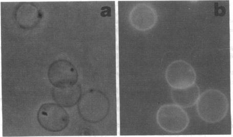
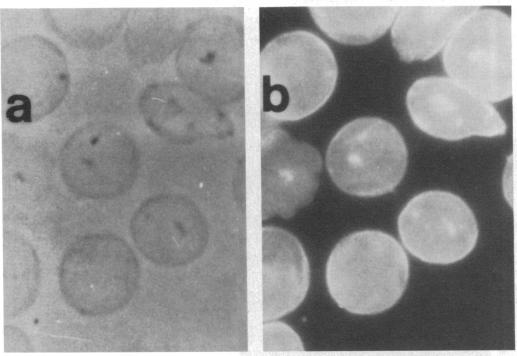
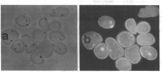
In earlier model studies we demonstrated that artificially denatured hemoglobin binds to and clusters the protein, band 3, in the plane of the erythrocyte membrane. To determine whether denatured hemoglobin also clusters band 3 in vivo, we have compared the locations of denatured hemoglobin aggregates (Heinz bodies) with band 3 in sickle cells using phase contrast and immunofluorescence microscopy. We report that where Heinz bodies are found associated with the cytoplasmic surface of the membrane, clusters of band 3 are usually colocalized within the membrane. In contrast, normal erythrocyte membranes and regions of sickle cell membranes devoid of Heinz bodies display an uninterrupted staining of band 3. Similarly, ankyrin and glycophorin are periodically seen to aggregate at Heinz body sites, but the degree of colocalization is lower than for band 3. These data demonstrate that the binding of denatured hemoglobin to the membrane forces a redistribution of several major membrane components.
在早期的模型研究中,我们证明了人工变性的血红蛋白在红细胞膜平面内与蛋白质带3结合并聚集。为了确定变性血红蛋白在体内是否也会使带3聚集,我们使用相差显微镜和免疫荧光显微镜比较了镰状细胞中变性血红蛋白聚集体(海因茨小体)与带3的位置。我们报告,在发现海因茨小体与膜的细胞质表面相关的地方,带3的聚集体通常在膜内共定位。相比之下,正常红细胞膜和没有海因茨小体的镰状细胞膜区域显示带3的连续染色。同样,锚蛋白和血型糖蛋白也会周期性地在海因茨小体部位聚集,但共定位程度低于带3。这些数据表明,变性血红蛋白与膜的结合迫使几种主要膜成分重新分布。