Omics Laboratory, Stanford University, Palo Alto, California, United States of America.
Department of Ophthalmology, Byers Eye Institute, Stanford University, Palo Alto, California, United States of America.
PLoS One. 2018 Feb 21;13(2):e0193250. doi: 10.1371/journal.pone.0193250. eCollection 2018.
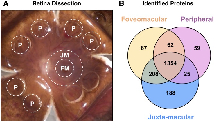
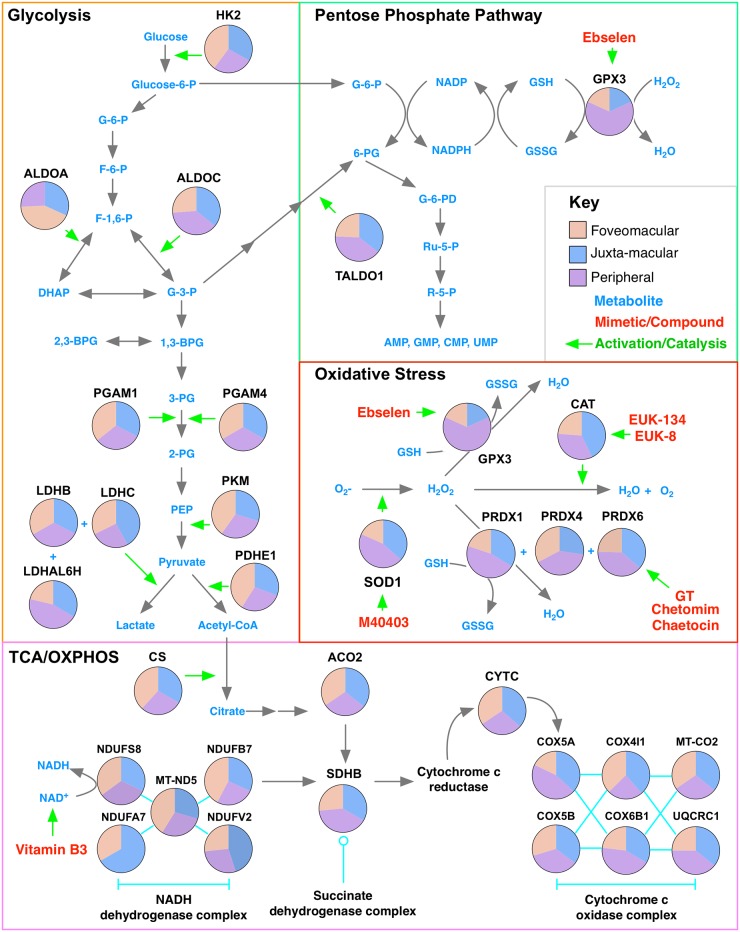
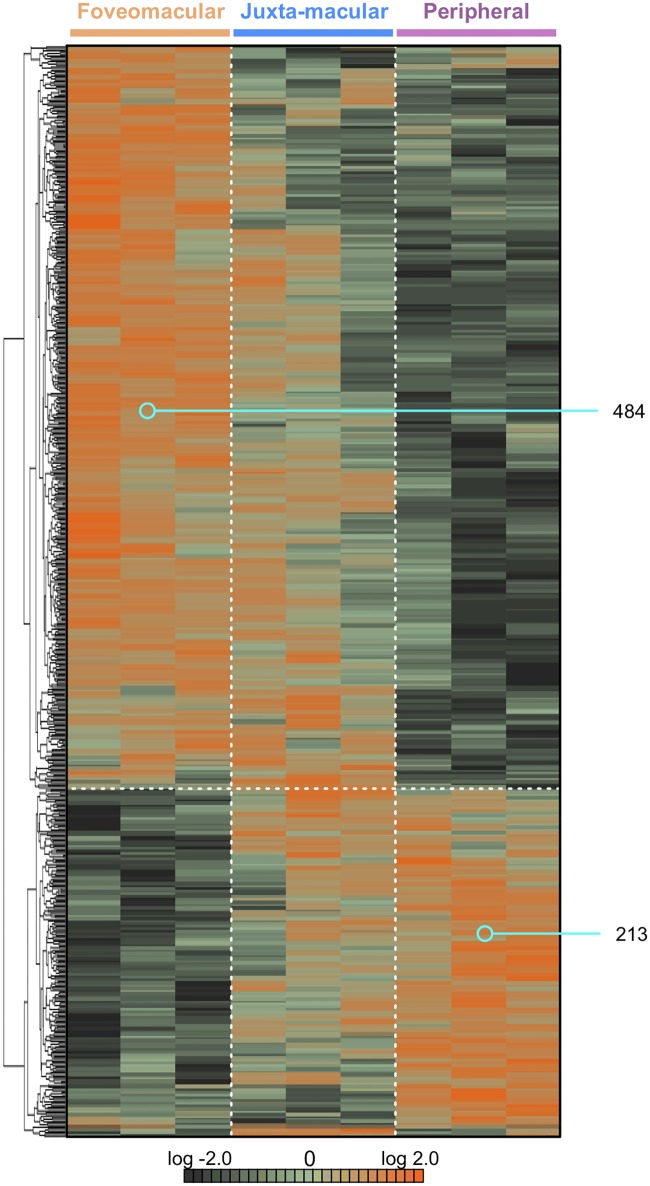
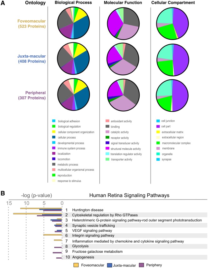
Differences in regional protein expression within the human retina may explain molecular predisposition of specific regions to ophthalmic diseases like age-related macular degeneration, cystoid macular edema, retinitis pigmentosa, and diabetic retinopathy. To quantify protein levels in the human retina and identify patterns of differentially-expressed proteins, we collected foveomacular, juxta-macular, and peripheral retina punch biopsies from healthy donor eyes and analyzed protein content by liquid chromatography-tandem mass spectrometry (LC-MS/MS). Protein expression was analyzed with 1-way ANOVA, gene ontology, pathway representation, and network analysis. We identified a mean of 1,974 proteins in the foveomacular retina, 1,999 in the juxta-macular retina, and 1,779 in the peripheral retina. Six hundred ninety-seven differentially-expressed proteins included those unique to and abundant in each anatomic region. Proteins with higher expression in each region include: heat-shock protein 90-alpha (HSP90AA1), and pyruvate kinase (PKM) in the foveomacular retina; vimentin (VIM) and fructose-bisphosphate aldolase C (ALDOC); and guanine nucleotide-binding protein subunit beta-1 (GNB1) and guanine nucleotide-binding protein subunit alpha-1 (GNAT1) in the peripheral retina. Pathway analysis identified downstream mediators of the integrin signaling pathway to be highly represented in the foveomacular region (P = 6.48 e-06). Metabolic pathways were differentially expressed among all retinal regions. Gene ontology analysis showed that proteins related to antioxidant activity were higher in the juxta-macular and the peripheral retina, but present in lower amounts in the foveomacular retina. Our proteomic analysis suggests that certain retinal regions are susceptible to different forms of metabolic and oxidative stress. The findings give mechanistic insight into retina function, reveal important molecular processes, and prioritize new pathways for therapeutic targeting.
人视网膜内区域蛋白表达的差异可能解释了特定区域对眼科疾病(如年龄相关性黄斑变性、囊样黄斑水肿、色素性视网膜炎和糖尿病性视网膜病变)的分子易感性。为了定量分析人视网膜中的蛋白质水平并确定差异表达蛋白的模式,我们从健康供体眼中采集了黄斑中心凹、近黄斑区和周边视网膜的活检,并通过液相色谱-串联质谱(LC-MS/MS)进行了蛋白质含量分析。使用单向方差分析、基因本体论、途径表示和网络分析对蛋白质表达进行了分析。我们在黄斑中心凹视网膜中鉴定出平均 1974 种蛋白质,在近黄斑区视网膜中鉴定出平均 1999 种蛋白质,在周边视网膜中鉴定出平均 1779 种蛋白质。697 种差异表达的蛋白质包括每个解剖区域特有的和丰富的蛋白质。每个区域表达较高的蛋白质包括:热休克蛋白 90-α(HSP90AA1)和丙酮酸激酶(PKM)在黄斑中心凹视网膜;波形蛋白(VIM)和果糖二磷酸醛缩酶 C(ALDOC);以及鸟嘌呤核苷酸结合蛋白亚基β-1(GNB1)和鸟嘌呤核苷酸结合蛋白亚基α-1(GNAT1)在周边视网膜。途径分析鉴定出整合素信号途径的下游介质在黄斑中心凹区域高度表达(P = 6.48 e-06)。代谢途径在所有视网膜区域中均有差异表达。基因本体论分析表明,与抗氧化活性相关的蛋白质在近黄斑区和周边视网膜中含量较高,但在黄斑中心凹视网膜中含量较低。我们的蛋白质组学分析表明,某些视网膜区域容易受到不同形式的代谢和氧化应激的影响。这些发现为视网膜功能提供了机制上的见解,揭示了重要的分子过程,并为治疗靶点确定了新的途径。