Swindle-Reilly Katelyn E, Paranjape Chinmay S, Miller Cheryl A
Department of Biomedical Engineering, Saint Louis University, 3507 Lindell Blvd., St. Louis, MO, 63103, USA.
Department of Chemical and Biomolecular Engineering, University of Pennsylvania, 220 South 33rd Street, Philadelphia, PA, 19104-6315, USA.
Prog Biomater. 2014 Feb 21;3(1):20. doi: 10.1007/s40204-014-0020-0.
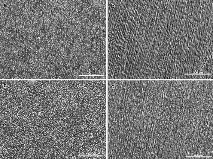
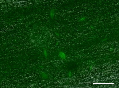
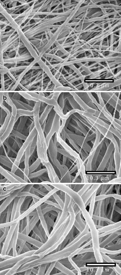
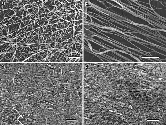
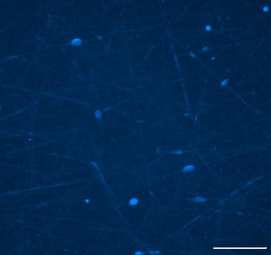
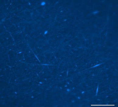
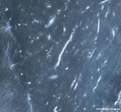
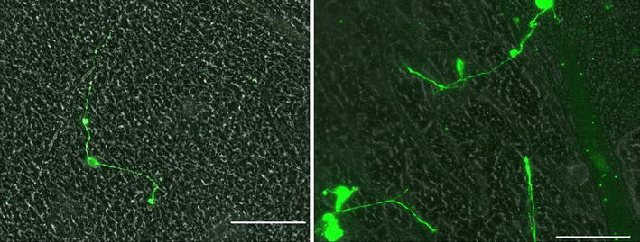
Peripheral nerve regeneration can be enhanced by chemical and mechanical cues for neurite growth. Aligned and randomly oriented electrospun nanofibers of poly(ε-caprolactone) (PCL) or a blend of PCL and elastin were fabricated to test their potential to provide contact guidance to embryonic chick dorsal root ganglia for peripheral nerve regeneration. Scanning electron microscopy was used to analyze the fiber diameter. Fiber diameter was found to be significantly smaller when elastin was incorporated into the scaffold (934 ± 58 nm for PCL and 519 ± 36 nm for PCL:elastin). After 24 h in culture, there was preferential cell attachment and neurite extension along the fibers of the elastin-containing scaffolds (average neurite extension 173.4 ± 20.7 μm), indicating that the presence of elastin promotes neurite outgrowth on electrospun scaffolds.
化学和机械性神经突生长线索可增强周围神经再生。制备了聚(ε-己内酯)(PCL)或PCL与弹性蛋白混合物的排列和随机取向的电纺纳米纤维,以测试它们为胚胎鸡背根神经节提供接触导向以促进周围神经再生的潜力。使用扫描电子显微镜分析纤维直径。当弹性蛋白掺入支架中时,发现纤维直径明显更小(PCL为934±58nm,PCL:弹性蛋白为519±36nm)。培养24小时后,细胞沿含弹性蛋白支架的纤维优先附着并发生神经突延伸(平均神经突延伸173.4±20.7μm),表明弹性蛋白的存在促进了电纺支架上的神经突生长。