Gordon Center for Medical Imaging, Division of Nuclear Medicine and Molecular Imaging, Department of Radiology, Massachusetts General Hospital / Harvard Medical School, Boston, MA, USA.
Athinoula A. Martinos Center for Biomedical Research, Department of Radiology, Massachusetts General Hospital / Harvard Medical School, Boston, MA, USA.
Brain Imaging Behav. 2019 Apr;13(2):333-344. doi: 10.1007/s11682-018-9847-7.
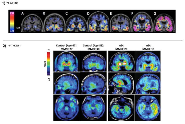
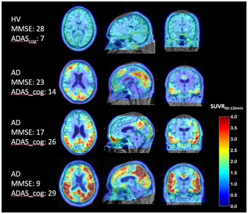
The two neuropathological hallmarks of Alzheimer's disease (AD) are amyloid-[Formula: see text] plaques and neurofibrillary tangles of tau protein. Fifteen years ago, Positron Emission Tomography (PET) with Pittsburgh Compound B (C-PiB) enabled selective in-vivo visualization of amyloid-[Formula: see text] plaque deposits and has since provided valuable information about the role of amyloid-[Formula: see text] deposition in AD. The progression of tau deposition has been shown to be highly associated with neuronal loss, neurodegeneration, and cognitive decline. Until recently it was not possible to visualize tau deposition in-vivo, but several tau PET tracers are now available in different stages of clinical development. To date, no tau tracer has been approved by the Food and Drug Administration for use in the evaluation of AD or other tauopathies, despite very active research efforts. In this paper we review the recent developments in tau PET imaging with a focus on in-vivo findings in AD and discuss the challenges associated with tau tracer development, the status of development and validation of different tau tracers, and the clinical information these provide.
阿尔茨海默病(AD)的两个神经病理学特征是淀粉样蛋白-β斑块和tau 蛋白的神经纤维缠结。15 年前,正电子发射断层扫描(PET)与匹兹堡化合物 B(C-PiB)相结合,能够选择性地在体内可视化淀粉样蛋白-β斑块沉积,并且自那时以来,已经提供了关于淀粉样蛋白-β沉积在 AD 中的作用的有价值的信息。tau 沉积的进展与神经元丧失、神经退行性变和认知能力下降高度相关。直到最近,tau 沉积才有可能在体内可视化,但现在有几种 tau PET 示踪剂处于不同的临床开发阶段。迄今为止,尽管研究工作非常活跃,但尚未有 tau 示踪剂获得美国食品和药物管理局批准用于 AD 或其他 tau 病的评估。本文综述了 tau PET 成像的最新进展,重点介绍 AD 中的体内发现,并讨论了 tau 示踪剂开发相关的挑战、不同 tau 示踪剂的开发和验证状态,以及这些示踪剂提供的临床信息。