Hur Kyu Yeon, Jun Ji Eun, Choi Young Ju, Lee Yong Ho, Kim Dae Jung, Park Seok Won, Huh Byung Wook, Lee Eun Jig, Jee Sun Ha, Huh Kap Bum, Choi Sung Hee
Division of Endocrinology and Metabolism, Department of Medicine, Samsung Medical Center, Sungkyunkwan University School of Medicine, Seoul, Korea.
Huh's Diabetes Center and 21st Century Diabetes and Vascular Research Institute, Seoul, Korea.
Diabetes Metab J. 2018 Feb;42(1):63-73. doi: 10.4093/dmj.2018.42.1.63.
The clinical utility of ankle-brachial index (ABI) is not clear in subjects with less severe or calcified vessel. Therefore, we investigated the usefulness of color Doppler ultrasonography for diagnosing peripheral artery disease (PAD) in type 2 diabetes mellitus (T2DM) subjects.
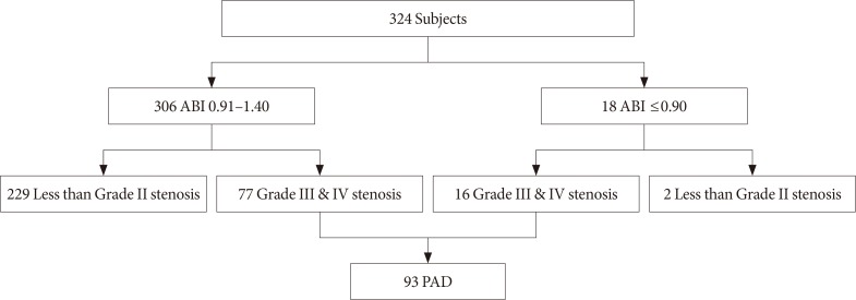
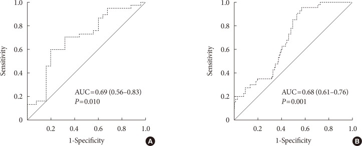
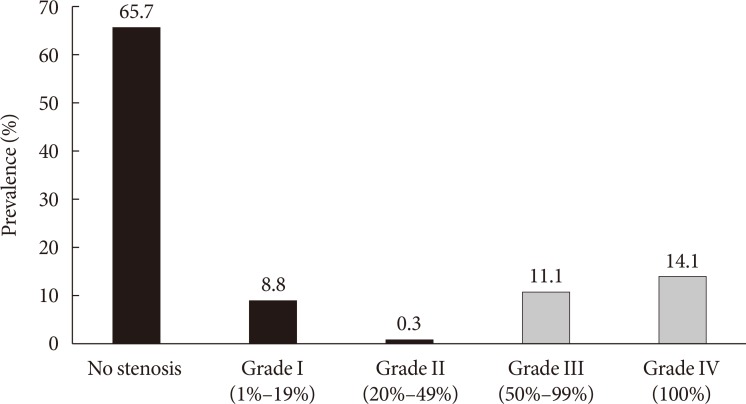
We analyzed 324 T2DM patients who concurrently underwent ABI and carotid intima-media thickness (CIMT) measurements and color Doppler ultrasonography from 2003 to 2006. The degree of stenosis in patients with PAD was determined according to Jager's criteria, and PAD was defined as grade III (50% to 99% stenosis) or IV stenosis (100% stenosis) by color Doppler ultrasonography. Logistic regression analysis and receiver operating characteristic curve analysis were performed to evaluate the risk factors for PAD in patients with ABI 0.91 to 1.40.
Among the 324 patients, 77 (23.8%) had ABI 0.91 to 1.40 but were diagnosed with PAD. Color Doppler ultrasonography demonstrated that suprapopliteal arterial stenosis, bilateral lesions, and multivessel involvement were less common in PAD patients with ABI 0.91 to 1.40 than in those with ABI ≤0.90. A multivariate logistic regression analysis demonstrated that older age, current smoking status, presence of leg symptoms, and high CIMT were significantly associated with the presence of PAD in patients with ABI 0.91 to 1.40 after adjusting for conventional risk factors. CIMT showed significant power in predicting the presence of PAD in patients with ABI 0.91 to 1.40.
Color Doppler ultrasonography is a useful tool for the detection of PAD in T2DM patients with ABI 0.91 to 1.40 but a high CIMT.
在血管病变不太严重或存在钙化的受试者中,踝臂指数(ABI)的临床应用价值尚不清楚。因此,我们研究了彩色多普勒超声在2型糖尿病(T2DM)患者外周动脉疾病(PAD)诊断中的作用。
我们分析了2003年至2006年期间同时接受ABI和颈动脉内膜中层厚度(CIMT)测量以及彩色多普勒超声检查的324例T2DM患者。根据耶格标准确定PAD患者的狭窄程度,彩色多普勒超声将PAD定义为III级(狭窄50%至99%)或IV级狭窄(狭窄100%)。进行逻辑回归分析和受试者工作特征曲线分析,以评估ABI为0.91至1.40的患者发生PAD的危险因素。
在这324例患者中,77例(23.8%)的ABI为0.91至1.40,但被诊断为PAD。彩色多普勒超声显示,ABI为0.91至1.40的PAD患者中,腘动脉上段动脉狭窄、双侧病变和多支血管受累的情况比ABI≤0.90的患者少见。多因素逻辑回归分析表明,在调整传统危险因素后,年龄较大、当前吸烟状态、腿部症状的存在以及高CIMT与ABI为0.91至1.40的患者发生PAD显著相关。CIMT在预测ABI为0.91至1.40的患者发生PAD方面显示出显著的效能。
彩色多普勒超声是检测ABI为0.91至1.40但CIMT较高的T2DM患者中PAD的有用工具。